Augmentation of hard tissue – an overview of regenerative techniques in dentistry
A scientific essay by Ralf Smeets, Germany
Correcting a qualitative or quantitative reduction in the bone bed before implantation can greatly improve the implant outcome. In addition to the gold standard of autologous bone grafting, the bone substitute materials currently available for this purpose can be subdivided into allogenic, xenogenic and alloplastic materials. Developments in the field of synthetic BSMs are constantly changing, and the selection of appropriate materials depends on the indication, availability and the individual treatment plan. The common goal of these BSMs is to achieve stable, long-term anchorage of the implants in the bone. A knowledge of the advantages and limitations of their principal properties in respect of osteoconduction, osteoinduction and osteogenesis, and the adequate selection, based on this knowledge, can ensure high-quality, evidence-based treatment.
Author: Ralf Smeets

Prof. Ralf Smeets
Dr. med. Dr. med. dent
Executive Senior Physician and Head of Research, University Medical Center Hamburg-Eppendorf, Head and Neurocenter of the Clinic and Outpatients for Oral and Maxillofacial Surgery, Hamburg, Germany Studied chemistry (specialist subject: macromolecular chemistry) and human medicine and dentistry at RWTH Aachen University. Surgeon specialized in oral and maxillofacial surgery. Dentist specialized in oral surgery Hans-von-Seemen Prize awarded by the German Association for Plastic and Restorative Surgery. Since 2011, Executive Senior Physician and Head of Research in the Clinic and Outpatients for Oral and Maxillofacial Surgery, University Medical Center Hamburg-Eppendorf. 2011 W2 University Professor of Maxillofacial and Oral Surgery in the Medical Faculty of Hamburg University.
Related products
Introduction
As well as the need for physical and mechanical integrity and stability in terms of adequate moldability during application, early load-bearing and ideal porosity, the focus of current research efforts is on the development of optimal biomolecular parameters in the area of osteogenesis (bone formation in the graft by osteoblasts), osteoconduction (creation of a scaffold for vascularization of the adjacent bone) and osteoinduction (differentiation of multipotent mesenchymal cells into osteoblasts, by proteins bound in the graft). In an ideal scenario, these parameters interact to achieve successful osteointegration of the BSM during the course of healing. In this case, the direct bonding of the bone substitute material to adjacent healthy bone without the formation of a separating layer of connective tissue can create the best possible basis for successful long-term implant provision. The implant-related indications determined by the dentist for the use of bone substitute materials predominantly involve horizontal and vertical bone defects of the alveolar ridge.
Autogenous bone substitute materials
Autogenous bone is considered to be the gold standard for hard tissue augmentation techniques. Apart from the physiological interaction between the aforementioned physical and mechanical parameters and outstanding performance in terms of osteoinduction, osteoconduction and osteogenesis, the use of autologous bone avoids complications such as contrary cellular and humoral immune reactions and transmission of diseases. However, the use of these grafts is associated with certain limitations: These include the need for a second intervention site with additional risks such as inflammation, pain or sensory disorders at the site of harvesting. Depending on the harvesting site (retromolar, mandibular symphysis, iliac crest), specific advantages and disadvantages in respect of the differing biological significance and the possible reconstruction volume will need to be taken into account1,2. Although autogenous bone is still considered to be the gold standard for long-term implant success, studies involving specific indications, such as lateral augmentation or the sinus lift, have shown that the use of bone substitute materials can achieve equivalent results. Moreover, the procedure of autogenous bone harvesting can significantly prolong operation time and the hospitalization rate in cases of bone harvesting of the iliac crest3,4.
Allogenic bone substitute materials
In order to counter the donor morbidity associated with the use of autogenous grafts, the use of allogenic grafts can be considered. Allogenic describes the transfer of the graft between genetically dissimilar individuals. Since the grafts count as medicinal products authorized e.g. by the Paul Ehrlich Institute in Germany or local accredited tissue banks, their use is guaranteed to be safe both for the therapist and patient. However, the risk of infection resulting from the transfer of biological tissue from human to human, as well as the risks of fractures or pseudarthroses associated with an allogenic graft, cannot be ruled out completely5. Through stringend donor screening and recovery protocols, as well as a highly controlled processing environment, the risks of disease transmission are countered at every step. To date there are no known cases of transmission triggered by processed, freeze-dried bone preparations in dental applications. Two distinct approaches can basically be adopted for processing allogenic BSMs: The material is freed from a given mineralized bone substance by decalcification in order to optimize the osteoinduction potential of the remaining growth factors in the collagen. This approach of demineralized bone matrix (DBM, also known as demineralized freeze-dried bone allograft, DFDBA), can be contrasted with the processing of the mineralized components of the donor tissue (mineralized freeze-dried bone allograft, FDBA). In this approach the tissue is freed of potentially infectious or immunologically active constituents. This leaves the natural bone structure with the trabecular matrix, with preservation of both the organic phase (collagen) and the mineral phase.
Since the physical and mechanical properties are equivalent to those of autogenous graft, allogenic BSM is considered to be comparable with autologous bone in respect of its osteoconductivity potential. One bone substitute material of human origin that is available in many European countries is the product maxgraft® from botiss biomaterials GmbH. This processed allogenic bone substitute is available in different standardized forms of granules, blocks, rings etc., or alternatively as an individually milled and shaped block graft, and can therefore be used for various indications. In case of large defects, a block-like matrix can be prepared from processed human donor bone, thus avoiding invasive harvesting of bone from the iliac crest or cranial areas. After organ donor selection and donor testing for infectious agents like HIV, HBV and HCV, the BSM undergoes chemical cleaning, preparation and sterilization. A solution tailored to the individual defect involves the use of a patient-specific bone block that is planned on the basis of imaging and then milled (maxgraft® bonebuilder, botiss biomaterials GmbH). As an individual allogenic graft solution, individual implants customized for the patient can be created by means of CBCT scanning in the CAD/CAM technique. These are constructed by botiss and then produced by the “Cells and Tissuebank Austria (CTBA)”. As a prefabricated ring structure (maxgraft® bonering), the allogenic graft supplied by the company botiss allows the bone augmentation and implantation to take place at the same time, avoiding the need for a second procedure. These grafts can be used in sinus floor augmentations or horizontal and vertical bone defects with simultaneous implant provision. The latest literature findings show results comparable with the use of demineralized allogenic BSM in sinus floor augmentations. The results of evidence-based studies also support the use of this type of BSM for alveolar ridge augmentations, intrabony defects and implant provision6-9.
Xenogenic bone substitute materials
In contrast with an allogenic BSM, xenogenic tissue is of animal or plant origin. In this context, BSMs of bovine origin are by far the most widely used materials. As with allogenic bone substitute materials, the matrix component is left in place during the processing of xenogenic grafts in order to control osteoconduction. The trabecular framework remains after thermal processing. Here, the osteoconductivity of the tissue as a scaffold for vessel formation and subsequent ingrowth of osteoblasts in the graft are particularly advantageous. As a result, biomolecular effective components are deactivated, leaving a matrix of hydroxyapatite ceramic. The lack of pathogen transmission due to this deactivation process is particularly beneficial. Additionaly, the immunological action can be considered to be low. Most of the commercially available products are designed to create a trabecular matrix for the formation of new bone. One example is cerabone® (botiss biomaterials GmbH), a material processed from bovine bone. Bone substitute materials obtained from equine bone or marine algae are also available. By far the most scientific evidence is available for the group of bovine xenogenic BSMs. Bovine bone material possesses physical and mechanical properties that are similar to those of human bone, thus comparable results can be achieved in terms of osteoconduction and vascularization.
Cerabone® is prepared by a unique high-temperature process that reliably removes potential infectious agents while at the same time preserving the natural cancellous bone structure. Due to the remaining trabecular support structure a maintenance of the original bone substitute volume can be achieved10, 11. The advantages of cerabone® include its high purity and volume maintenance, an interconnecting pore structure and minimization of the infection risk by its processing12. A feature common to all BSMs of bovine origin is the comparatively low remodeling potential during healing. As a result, the bone substitute material remains in situ as a slowly-resorbable osteoconductive scaffold.
Alloplastic bone substitute materials
Alloplastic bone substitute consists of synthetically produced materials. By imitating the trabecular bone matrix of human bone, the synthetic material possesses similar osteoconductive and osteointegrative capabilities. Depending on the starting material, the alloplastic materials can be subdivided as follows: ceramics (tricalcium phosphate, hydroxyapatite, bioglasses, glass ionomers), polymers (polymethyl methacrylate, polylactides/polyglycolides and other copolymers), cements (calcium phosphate cements) and metals (titanium). Durability and resorbability are particularly important when selecting these materials.
Ceramics
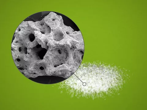
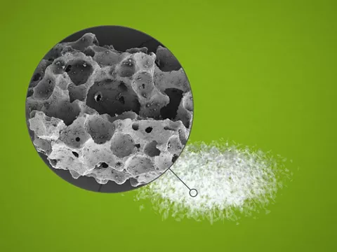
Alloplastic hydroxyapatite ceramics [Ca10(PO4)6(OH)2] are the most common representatives of this group. The sintering of calcium phosphates (hydroxyapatit (HA), alpha-tricalcium phosphate (α-TCP) and beta-tricalcium phosphate (β-TCP)) produces a biocompatible, non-immunogenic and a slowly-resorbable matrix with osteoconductive and osteointegrative properties13-16. Since hydroxyapatite in crystalline form provides the basis for the hard substance of human bone, making up around 40 % of its content, ceramics of this type are likely candidates for use in bone replacement. Innovative sintering techniques can create the conditions that are favorable for osseointegration and biodegradation. Here, pore diameters of 150–600 μm are considered to be ideal for these purposes14.
A fully synthetic commercially available representative of this group is maxresorb® (botiss biomaterials GmbH). Consisting of 60% hydroxyapatite (HA) and 40% beta-tricalcium phosphate (ß-TCP), maxresorb® possesses a uniform interconnecting pore structure thanks to the standardized manufacturing process. With pore sizes ranging from 200 to 800 μm and an overall porosity of ~80%, it provides a suitable scaffold for vascularization and cell migration.18
Indications for the dentist in practice
Returning to the initially mentioned indications for the use of bone substitute materials in implantology, the following recommendations can be made on the basis of the latest guideline on “Implantology-related indications for the use of bone substitute materials”:
Dehiscence defect (intrabony defect): In most cases, the use of BSMs resulted in complete defect regeneration.
Horizontal/vertical defects: The use of BSMs produced horizontal (3.6–5.6 mm) and vertical (2.0–5.6 mm) dimensional gains after 6 months with values of less than 5 % for augmentation and implant losses. Xenogenic BSMs were superior to allogenic grafts in respect of the newly-formed bone.
Sinus floor elevation: Xenogenic BSMs (95.6 %) and allogenic BSMs (93.3 %) were superior to alloplastic materials (81 %).
External sinus floor elevation: The use of BSMs resulted in a cumulative implant survival rate of almost 97 %. Autologous bone is inferior to particulate grafts.
Internal sinus floor elevation: The use of BSMs resulted in implant survival rates of 94.8-100 %. No general recommendation was issued for the use of BSMs in this context.
It can therefore be assumed that the use of alloplastic bone substitute materials can achieve very good results particularly in the management of alveolar dehiscence defects (up to a height of 8mm) and also in sinus floor elevations. Augmentations with purely allogenic grafts in the whole vertical-horizontal situation and for fairly large defects should be handled more critically. Current recommendations point to the benefits of additive supplementation with allogenic BSMs in procedures with autologous bone substitute materials. Extensive defects should generally be reconstructed with large-pore BSMs, ideally with pore diameters in the range of 150 to 600 μm, in order to facilitate better neovascularization, cell permeation and osteoconduction.
Summary
Depending on the individual indication, the dentist can currently choose from a wide range of evidence-based bone substitute materials. Both natural and allogenic BSMs can basically be used, either on their own or in addition to endogenous bone, for the reconstruction of jaw defects. To this end, the advantages and disadvantages and possible contraindications of the respective material groups will need to be assessed and used on a case-by-case basis. Before planning the treatment, the dentist must conduct an individual, patient-oriented evaluation of the osteogenic efficiency and relative risks for the patient arising from the use of allogenic or xenogenic bone substitute materials. Given the complexity and diversity of the subject of “bone substitute materials”, careful and comprehensive briefing of the patient is extremely important before any augmentation measure, regardless of the material that is ultimately used. Relative and, where applicable, absolute contraindications to the use of alloplastic materials are an immunocompromised patient (immune deficiency, interleukin-1 polymorphisms), generally poor oral hygiene (dental status not worth preserving, severe chronic periodontitis), treatment with drugs that inhibit bone resorption (bisphosphonates) and previous oral-maxillofacial radiotherapy.
Finally, it should be stressed that the correct technique is of crucial importance when using the respective bone substitute material. Apart from the choice of suitable bone substitute material, key factors for long-term success of the implant are the membrane application, the appropriate incisions and the soft tissue management.