The Straumann® PURE Ceramic Implant and the digital prosthetic workflow
A clinical case report by Ulises Calderon/Stefan P. Hicklin, Switzerland
The introduction of oxide ceramics in dentistry provided new treatment options for patients and clinicians. To date, zirconia has shown superior biomechanical properties compared to other oxide ceramics. Since it was introduced in dentistry, zirconia has been used as a framework material for all ceramic crowns and fixed dental prostheses (FDP), as well as for implant abutments. Because of its material properties and tooth-like color, zirconia is also the material of choice for dental implants nowadays. Moreover, human studies have demonstrated a reduced bacterial adhesion on zirconia compared to titanium, as well as fewer inflammatory cells in the peri-implant soft tissue of zirconia compared to titanium. These findings suggest that less periimplantitis may occur around zirconia implants compared to titanium implants. In a recent systematic review (Hashim et al 2016), the overall survival rate of one- and two-piece zirconia implants after 1 year of function was 92% (95% CI 87-95). According to this review, it seems that zirconia implants could serve as a metal-free alternative to titanium implants. On the other hand, it has to be pointed out that there is still a lack of data regarding the long-term performance of zirconia implants. Further clinical studies are needed in order to obtain more data on the long-term outcome of zirconia implants. In this context, case reports are also valuable for identifying the risk factors for technical and biological complications.
Initial situation
A 69-year old patient attended her scheduled maintenance appointment at the University Clinics of Dental Medicine in Geneva (Division of Fixed Prosthodontics and Biomaterials) in February 2014. The patient was healthy and a non-smoker. During the routine examination she complained of a slightly painful sensation at tooth 35. The tooth had been endodontically treated and restored with a crown two years ago. The clinical evaluation confirmed the slight pain on percussion of tooth 35. Furthermore, the tooth showed increased mobility of grade 2 and a localized probing pocket depth of 7mm. A periapical radiograph to complete the examination showed a radiolucency mesial to the root (Fig. 1). A presumptive diagnosis of vertical root fracture was made.
Treatment planning
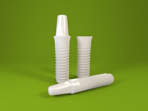
An exploratory surgery was planned in the Division of Periodontology (University of Geneva). If the presumptive diagnosis was confirmed, the plan was to extract tooth 35 and perform a ridge preservation procedure. After the bone healing period, late implant placement of a Straumann® PURE Ceramic Implant 4.1 diameter on position of 35 was planned. After successful osseointegration, the implant was to be restored with a hybrid ceramic (VITA Enamic®, VITA Zahnfabrik, Bad Säckingen, Germany) crown using an intra-oral optical impression and CAD/CAM technology to follow a digital workflow.
Surgical procedure
The exploratory surgery was performed in May 2014 and confirmed the suspected diagnosis of a vertical root fracture of tooth 35. As planned, the tooth was extracted (Fig. 2) and a xenogenic bone substitute was used to perform a ridge preservation procedure. No provisional prosthesis was inserted to replace the missing tooth. Ten months later, because the patient had spent some months abroad, a well maintained and completely healed ridge was observed clinically (Fig. 3). Alginate impressions of the situation were made and a wax-up of tooth 35 was fabricated on the plaster models. Based on the wax-up, the dental technician manufactured a conventional surgical guide. On the day of the surgery, the patient was pre-medicated with 750mg amoxicillin and 600mg ibuprofen. Under local anesthesia, a mid-crestal incision with a distal vertical releasing incision was performed, and a full thickness flap was elevated (Fig. 4). Because the patient already had an implant replacing tooth number 36, the design of the incision respected the mesial mucosa around this implant. The conventional surgical stent was placed, and the implant bed was prepared according to the manufacturer’s instructions (Fig. 5). The preparation and correct vertical positioning for a monotype zirconium dioxide ceramic implant with an endosteal diameter of 4.1mm were achieved with the help of the specific implant indicators (Figs. 6-8). A Straumann® PURE Ceramic Implant with a diameter of 4.1mm, length of 8mm and an abutment height of 4mm was placed with good primary stability (Figs. 9-11). A xenograft and a collagen membrane were used to augment the small buccal bone defect and enhance the vestibular ridge contour for a more natural appearance of the final crown. The respective protective cap was placed on the abutment part of the implant. The flap was repositioned and adapted with a 5.0 ePTFE non-absorbable monofilament suture. Antibiotics, pain killers and chlorhexidine rinse mount were prescribed. The sutures were removed 10 days after implant placement.
Prosthetic procedure
Healing was uneventful and, after 8 weeks, clinical examination revealed healthy and stable tissues. During the same visit the protective cap was modified (Figs. 12 – 13) with composite on the mesial aspect in order to condition the soft tissue in this area and allow good registration of the prosthetic shoulder with an intraoral scanner. One week later the intraoral optical impression was taken (Cerec® Omnicam, Dentsply Sirona, Germany), and a single crown was designed chair-side using CAD Software (CEREC® software 4.3, Dentsply Sirona, Germany) (Figs. 14 – 16). A hybrid ceramic (VITA Enamic®, VITA, Bad Säckingen, Germany) was chosen as a restorative material because it was assumed that its load-bearing capacity was increased, thus leading to less chipping and greater chewing comfort for the patient. The crown was milled in the lab using a 3-axis CAM machine (CEREC® MC X, Dentsply Sirona, Germany). After the try-in of the crown (Fig. 17), minor occlusal adjustments were necessary. To improve the shade of the final crown, staining products were used in the dental laboratory according to the manufacturer’s recommendations (VITA Enamic® Stains, Vita, Bad Säckingen, Germany). One day after the try-in, the crown was already finished and could be cemented onto the implant (Fig. 18). The internal part of the crown was etched with 5% hydrofluoric acid (IPS Ceramic Etching gel, Ivoclar Vivadent, Schaan, Liechtenstein) for 60 seconds, then cleaned with water and, in an ultrasonic bath, with alcohol for 5 minutes. A universal primer (Monobond Plus®, Ivoclar Vivadent, Schaan, Liechtenstein) was applied to the etched surface of the crown. The mucosa around the implant shoulder was displaced using a retraction cord to avoid cement excess at the level of the implant shoulder (Fig. 19). The abutment part of the Straumann® PURE Ceramic Implant was cleaned and pretreated for adhesive cementation. The same universal primer used for the crown (Monobond Plus®, Ivoclar Vivadent, Schaan, Liechtenstein) was applied to the abutment surface. Self-curing MDP containing resin cement (Panavia 21 TC, Kuraray, Japan) was used to cement the crown onto the implant abutment part. The excess of cement and the retraction cord were carefully removed and the occlusion was rechecked. A periapical radiograph was taken (Fig. 20) to verify the absence of residual cement around the implant. Two control visits were arranged at 3 days and 10 days after cementation (Figs. 21 – 22). No further adjustments of the crown were necessary, and the patient was very comfortable with the final reconstruction.
Final results
Maintenance and control appointments were arranged at 6 months and 1 year after implant placement. Clinically, the crown on the implant was still functional, and no technical complications were recorded at these two time points (Figs. 23 – 24). The soft tissue around the implant 35 remained healthy. A periapical radiograph was taken at the last control visit, 1 year after implant placement. Normal bone remodeling around the implant was observed, and the bone level stabilized around the border with the rough implant surface (Fig. 25). The patient was very satisfied with the treatment outcome in respect of both function and esthetics.
Conclusion
After one year of function, no biological or technical complications were apparent. The treatment option with this type of zirconia implant and restoration with a hybrid ceramic crown in the described indication seems to be a valid alternative to titanium implants. The soft tissue around the implant remained stable over time, which shows the excellent biocompatibility of the zirconia ceramic. It should be mentioned that the vertical implant position is a crucial factor for success. As it is a one-piece implant, the reconstruction has to be cemented, which involves the risk of cement excess around the implant, especially if the implant shoulder is placed too deep beneath the mucosa.


