Straumann® keramische
Gingivaformer

Der erste Schritt für eine harmonische Weichgewebsheilung.

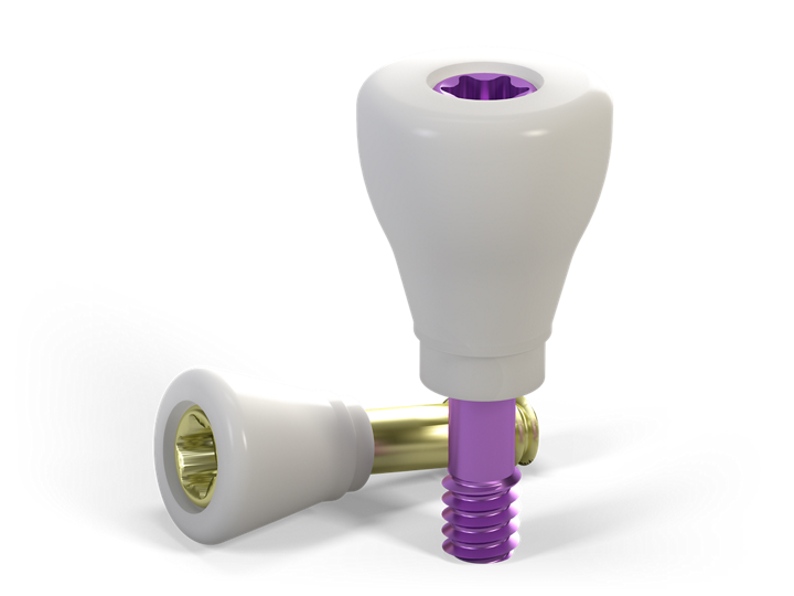
Die Straumann® Ceramic Gingivaformer begünstigen die Bildung der epithelialen Attachments und schaffen so eine gesunde periimplantäre Umgebung. Das bewährte Material Zirkondioxid reduziert Plaqueansammlungen und bewirkt ein exzellentes Weichgewebsverhalten schon ab dem Tag des chirurgischen Verfahrens.

Wichtige Indikationen
Einzelzahnversorgung
Fixierung
Material
ZrO2
Workflow
Klassisch
Implantatsysteme
BL | BLT
Prothetikverbindungen
NC, RC
Begünstigt die Bildung des epithelialen Attachments

Im Vergleich mit Titan tragen Zirkondioxidmaterialien allgemein zu einer verbesserten Bildung der epithelialen Attachments bei. Die periimplantäre Gewebedurchblutung ist mit der Gewebedurchblutung um den natürlichen Zahn vergleichbar.1–2

Entwickelt für eine gesunde periimplantäre Umgebung.

Dank der glatteren Oberfläche weniger Plaqueansammlung auf Zirkondioxid verglichen mit Titan.2–3,8–9 

Anwenderfreundlich

Aspirationsschutz dank integrierter Schraube.
Farbkodiert zur eindeutigen Identifizierung der zugehörigen prothetischen Plattform.

Ästhetische Ergebnisse schon am Tag des chirurgischen Verfahrens

Keramik-Sekundärteile für die Einheilphase. 
Definitive Versorgung unter Verwendung der Straumann® CARES® Keramikoptionen.

Broschüren und Videos

Sie suchen weitere Informationen? Sie finden sie in der Mediathek.

Sehenswerte Beiträge auf youTooth

Kontakt

Anrede
Datennutzungsvereinbarung*

Bitte beachten Sie unsere Datenschutzhinweise.

Literatur

1 Kajiwara N, Masaki C, Mukaibo T, Kondo Y, Nakamoto T, Hosokawa R (2015). Soft tissue biological response to zirconia and metal implant abutments compared with natural tooth: microcirculation monitoring as a novel bioindicator. Implant Dent 24(1):37-41. 2 Degidi M, Artese L, Scarano A, Perrotti V, Gehrke P, Piattelli A. Inflammatory infiltrate, microvessel density, nitric oxide synthase expression, vascular endothelial growth factor expression, and proliferative activity in peri-implant soft tissues around titanium and zirconium oxide healing caps. J Periodontol. 2006 Jan;77(1):73-80. 3 Liñares A, Grize L, Muñoz F, Pippenger BE, Dard M, Domken O, Blanco-Carrión J. Histological assessment of hard and soft tissues surrounding a novel ceramic implant: a pilot study in the minipig. J Clin Periodontol. 2016 Jun;43(6):538-46.  4 Erbshäuser M., Zirconium dioxide dental implants in single-tooth gaps – An alternative to titanium? (Article in German). Implantologie Journal 11 | 2015, 32-36. 5 Welander M, Abrahamsson I, Berglundh T. The mucosal barrier at implant abutments of different materials. Clin Oral Implants Res. 2008 Jul;19(7):635-41. 6 Tetè S, Mastrangelo F, Bianchi A, Zizzari V, Scarano A. Collagen fiber orientation around machined titanium and zirconia dental implant necks: an animal study. Int J Oral Maxillofac Implants. 2009 Jan-Feb;24(1):52-8. 7 De Medeiros RA, Vechiato-Filho AJ, Pellizzer EP, Mazaro JV, dos Santos DM, Goiato MC (2013). Analysis of the peri-implant soft tissues in contact with zirconia abutments: an evidence-based literature review. J Contemp Dent Pract 14(3):567- 572. 8 S. Röhling, M. Astasov-Frauenhoffer, I. Hauser-Gerspach, O. Braissant, H. Engelhardt, T. Waltimo, M. Gahlert. In vitro biofilm formation on commercially available machined and micro-roughened titanium and zirconia implant surfaces. Clin. Oral Impl. Res. 26 (Suppl. 12), 2015. 9 Scarano A, Piattelli M, Caputi S, Favero GA, Piattelli A. Bacterial adhesion on commercially pure titanium and zirconium oxide disks: an in vivo human study. J Periodontol. 2004 Feb;75(2):292-6.