Fully validated digital workflow with Straumann® CARES® Digital Solution and coDiagnostiX®
A clinical case report by Ferit Bayram, Turkey
Immediate loading of dental implants with a fully digital workflow offers several advantages, for example, reduced chair time for both the surgical and prosthodontic procedures. Digital prosthetic design enables future temporary crowns to be planned and prepared with optimal margins, occlusion and contacts with neighboring teeth. Evaluating the bone quality and quantity, selecting the appropriate implant and its position are crucial factors for the future crown. Fully guided surgery ensures prosthetically driven implant placement with an ideal position.
This report presents a successful restoration of a compromised single missing tooth in the esthetic zone, using Straumann® CARES® Visual and coDiagnostiX®. A fully digital workflow with digital planning, guided surgery, CADCAM production of a temporary crown and immediate loading meets the patient´s high esthetic expectations on the same day of operation.
This case was part of the Solution Driven Learning (SDL) education program at Marmara University in Istanbul, Turkey. SDL is an advanced education format that encourages students to identify, analyze and solve an open-ended problem by themselves. Mentoring plays a key role in this student-centered approach. Over a period of three days, six postgraduate students received cases with a single missing tooth in the esthetic zone. With the previously trained coDiagnostiX® and CARES® Visual, they restored the cases and presented the results in front of their Marmara University peers. Dr. Bayram’s case was selected for publication through an open competition. Dr. Gökhan Göçmen, Dr. Şükrü Can Akmansoy, Dr. Zeliha Şanıvar, Dr. Seda Keban and Dr. Gökhan Gedikli attended the program together with Dr. Bayram, and Prof. Yasemin Özkan and Assoc. Prof. Umut Arslan mentored the attendees. Straumann Turkey and the Medical Education department at Straumann HQ supported the whole program.
About Ferit Bayram

Ferit Bayram
DDS, PhD
Ferit Bayram received his DDS and PhD degrees from Marmara University, Faculty of Dentistry, Istanbul. He is currently practicing at Marmara University in the Department of Oral and Maxillofacial Surgery, and continues his academic work, which focuses on oral surgery, advanced implant surgery and orthognathic surgery.

Initial situation
A 20-year-old female patient was referred to our department for a missing #25 (Fig. 1). She had no known allergies and no medication history. Her chief complaint was poor esthetics due to the missing premolar, and her esthetic expectations were high. Intraoral examination revealed a medium-thick gingival phenotype, good periodontal status and good oral hygiene with enough restorative space to place an implant-supported prosthesis (Figs. 2-3).
3D radiographic imaging with Cone Beam Computed Tomography (CBCT) revealed a sufficient buccolingual width of 5.5 mm with an appropriate vertical dimension of 10 mm, requiring neither simultaneous nor staged bone grafting (Fig. 4). Bone density was type II according to the Leckholm and Zarb classification. The SAC assessment tool was used to identify the degree of complexity and potential risks involved in this case. Because of an immediate loading protocol and the other modifiers, this case was determined as advanced (Figs. 5-6).
Treatment planning
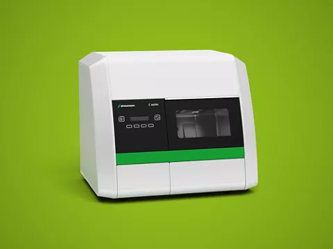
After discussing different treatment alternatives, we decided on immediate loading with a fully digital workflow to deliver a digitally produced temporary crown on the same day of surgery. For the education program, diagnostic cast models were prepared in advance by the mentors, and scanned STL data was given to the mentees (Figs. 7-8). These STL files were imported into the coDiagnostiX® software, where DICOM images from the patient’s CBCT were segmented and merged. After segmenting and landmarking important anatomical points, prosthetically driven implantation was planned, taking account of the available bone volume and the soft tissue profile, using coDiagnostiX®. A Straumann® Bone Level, Ø 3.3 mm, Narrow Neck, 10 mm length, Roxolid®, SLActive® implant was selected (Fig. 9). The surgical guide was designed with a sleeve of 5.0 mm in diameter and 5.0 mm in height. This guide design was sent to the lab and printed using the Straumann® CARES® P30 printer.
Surgical procedure
The implant bed was prepared with Straumann® Guided Surgery using the printed surgical guide. Proper fitting of the surgical guide, stability and bilaterally balanced passive seating were confirmed before the surgery (Fig. 10). First, local anesthesia was administered (Ultracaine®, Hoechst Marion Roussel, 40 mg articaine hydrochloride and 0.012 mg epinephrine hydrochloride). Following the surgical protocol, the mucosa was removed with a Mucosa punch (Figs. 11-12), and then a Milling Cutter, Ø 2.8 mm guided, was used to even the alveolar bone. Using a BLT Guided Pilot drill Ø 2.2 mm and a BLT Guided drill Ø 2.8 mm with a Straumann® drill handle with three dots' side, the implant bed was prepared under copious sterile saline irrigation (Figs. 13-14). The osteotomy was completed with a Ø 3.3 mm guided profile drill in combination with a C-handle H6. The Straumann® Bone Level, Ø 3.3 mm, Narrow Neck, length 10 mm, Roxolid®, SLActive® implant was placed, achieving a final torque of 50 N.cm (Fig. 15). After removal of the guided implant transfer piece, the ISQ value of the implant was measured with the Osstell device. This showed an ISQ of 74, a value that allowed immediate loading (Figs. 16-17). No suturing was necessary thanks to the flapless approach.
The patient was prescribed 1000 mg amoxicillin/clavulanic acid twice daily for five days (Augmentin®, Glaxo Smith Kline), 50 mg diclofenac (Cataflam®, Novartis) every 8 hours in case of pain, and chlorhexidine (Klorhex®, Drogsan) for gargling. The patient reported no discomfort after surgery.
Prosthetic procedure
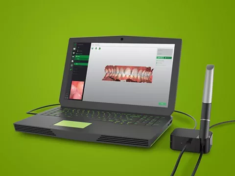
A digital impression was obtained with an intraoral scanner (Trios, 3Shape), initially from the upper jaw, with and without a scanbody connected to the implant (Fig. 18). The lower jaw and the patient’s bite were scanned, and the data was transferred as .stl files to Straumann® CARES® Visual, where the temporary prosthesis was designed (Fig. 19). The designed screw-retained temporary crown was sent to the third-party dental laboratory and manufactured using Sigmadent PMMA blocks. Self-adhesive resin cement (Multilink Speed, Ivoclar Vivadent, Schaan, Liechtenstein) was applied to the Variobase®, and the temporary restoration was bonded extra-orally. After polymerization of the resin cement, excess cement was removed from the Variobase®-temporary crown complex, and the margin of the was polished. The temporary crown was placed on the implant and tightened by hand (Figs. 20-21). After the adjustment of the contouring and the occlusion of the crown, a small cotton plug was inserted into the screw access channel. The occlusion required only minor adaptations due to accurate digital planning. The rest of the channel was filled with a temporary filling material. The provisional implant restoration was worn for three weeks to allow proper re-contouring of the soft tissue complex (Fig. 22). After three months, the patient was still content with the esthetics and function of the provisional restoration and ready for the final restoration (Figs. 23-24).
Treatment outcome
A fully digital workflow—digital planning, guided surgery, digital impression taking, computer-assisted design and manufacture (CADCAM) of a temporary crown for the immediate loading on the day of surgery—has three main advantages, if properly selected, planned and applied. Reduced chair-time, using the whole potential anatomical and biological conditions and presenting the patient with a perfectly acceptable prosthetic rehabilitation in one day. The total treatment time and the number of surgical procedures are also reduced.
The expectation of a successful outcome with this treatment protocol relies on two basic principles: proper indication and sufficient primary stability. Factors affecting primary stability, such as implant design, drilling protocol, bone density, and morphology, should be taken into consideration before deciding to proceed with this treatment protocol.