Restoration of a lateral maxillar incisor, missing due to aplasia
A clinical case report by Florian Thieringer, Switzerland
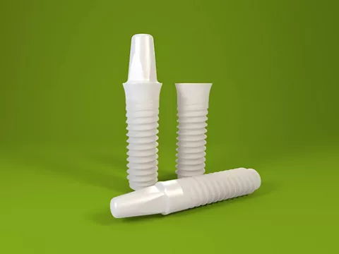
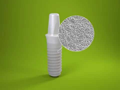
"This one-piece 3.3 mm diameter-reduced Straumann PURE Ceramic Implant is what we have been waiting for. The 3.3 mm implant extends the therapeutic possibilities, particularly in situations where the diameter of the 4.1 mm ceramic implant is too wide or in the esthetically demanding region of the anterior teeth. This exhibits the same outstanding properties as the 4.1 mm Straumann
PURE Ceramic Implant with respect to surface and biocompatibility. The micro- and macro-rough surface of the ZLA
has been subjected to experimental and clinical tests and guarantees reliable osseous ingrowth, as with the Straumann Titanium implants with SLA
surface. In addition, the new 3.3 mm ceramic implant exhibits outstanding fracture strength even at the lower diameter. Thus it is an ideal tooth-coloured implant for reliable and esthetic care when space is limited." Dr. Florian Thieringer
Author: Florian Thinger

Florian Thinger
Dr. med. et med. dent., MHBA
Specialist in Oral and Cranio-Maxillofacial Surgery, Consultant at the Department of Oral and Maxillofacial Surgery, University Hospital Basel, Switzerland. Medical Degree of the Munich Technical University, Dental Degree of the Ludwig-Maximilians-University Munich, Master of Health Business Administration of the FA University Erlangen-Nuremberg, Germany. Member of the International Team of Implantology (ITI).

Initial situation
A 22-year old female patient suffered aplasia of both lateral maxillar incisors. After orthodontic treatment, the local dentist filled the intermediate spaces in regions 12 and 22 with two Implants. The patient came to our clinic with advanced peri-implantitis on the implant in region 22 (Figs. 1-2). An impression was taken of the maxilla and mandible. Then a removable single tooth clamp temporary prosthesis was fabricated pre-operatively, supported occlusally on the neighbouring teeth. This was to fill the space that arose after explantation of the implant.
Surgical procedure
The implant in region 22 was explanted and the tissue sections modified by inflammation were carefully curetted. As suggested by the X-rays, the operation found a significant multiwalled bone defect (Figs. 2-3). Eight weeks after explantation, the defect region was augmented with an autologous bone block from the mandibular angle as well as bone substitute material (botiss cerabone®) and a collagen membrane (botiss Jason® membrane). Six months after bone augmentation, a one-piece diameter-reduced ceramic implant was implanted (diameter: 3.3/length: 10 mm). Because of the narrowness of the mesio-distal spaces in region 22, only a diameter-reduced implant could be used here. On the buccal side, autologous bone chips from the zygmomatic bone region were harvested (Figs. 4-5). The healing process was normal(Fig. 6). The implant was exposed to the oral cavity and healed without inflammation under the protection of the pre-operatively fabricated temporary clamp prosthesis.
Prosthetic procedure
To form the gingival soft tissue, a single temporary tooth restoration was fabricated from composite on a Straumann temporary coping. Optimal soft tissue formation was then ensured using a CAD long-term temporary restoration (Fig. 7). After approx. 16 weeks, the final impression was taken for a full ceramic crown (Fig. 8). The postoperative X-ray shows the correction positioning of the implant in the maxilla bone (Fig. 9).
Conclusion
It was possible to treat the highly esthetically challenging situation with limited space and significant bone deficit well using a gradual treatment concept. The choice of a reduced-diameter 3.3 mm Straumann one-piece ceramic implant with macro- and micro-rough surface and optimal biocompatibility ensured not only treatment success but also met the desire of the patient for a tooth-colored, completely metal-free solution, with a dark titanium implant showing through the gingiva on the opposite side. The measures for GBR were consistent with the usual approach for titanium implants.
The treatment described in this article was performed with substantial contributions from Dr. Michael Gahlert, Munich (Germany) and Dr. Stefan Röhling, Basel (Switzerland).