Single immediate implant and immediate screwed provisional prostheses using Straumann® BLX implants
A clinical case report by Juan Blanco, Spain; Iria Darriba, Spain; Javier Pérez, Spain
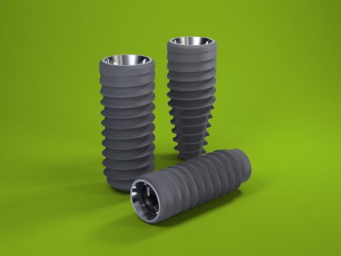
Scientific evidence (pre-clinical and clinical studies) in recent decades has shown that, in ideal situations, immediate implant placement with immediate loading has survival and success rates similar to those for delayed protocols. In the case reported here we used the new Straumann® implant design: BLX, with the SRA (one abutment-one time protocol) and with immediate provisional screwed prostheses. Based on our research, this treatment protocol using a 2.5 mm abutment installed at the time of surgery, could be an ideal solution for preserving peri-implant crestal bone.
Initial situation
A 45-year-old male patient came to our clinic to restore the two fractured upper second premolars (15 and 25) (Fig. 1). His personal history was unremarkable; he was a non-smoker with no systemic diseases (ASA I) or periodontal disease. After clinical and radiological analyses, an immediate implant placement and loading protocol was decided.
Treatment planning
Due to the systemic and local healthy situation, the immediate protocol was planned. Clinical examination of both premolars revealed no gingival recession, adequate width and thickness of keratinized gingiva (thick biotype) (Fig. 1), a shallow probing pocket depth and no bleeding on probing. From a radiological diagnosis we confirmed an integral and thick buccal bone plate, no radiolucency at the apex, good bone availability and alignment between the roots and the alveolar crest (Figs. 2-3). After the prosthetic analyses (casts, wax-up and smile design), we proceeded to design and manufacture two provisional crowns and create a surgical stent. With all this information we planned to use two 3.75 mm x 12 mm Straumann® BLX, Roxolid®, SLActive® implants with two 2.5 mm SRAs height.
Surgical procedure
After local anesthesia, we very gently extracted the two premolars (the right one with a flap approach and the left with flapless approach) and thoroughly cleaned both alveoli with curettes and saline (Fig. 4). Next, following the manufacture guidelines we proceeded to insert the implants. We paid particular attention to achieving good primary stability (at least 20 Ncm). We filled the gap between the implants and buccal bone plate with Straumann® XenoGraft (deproteinized bovine bone mineral), which is a slowly-resorbing bone substitute (Figs. 5-6). Finally, the two SRAs were screwed on top of the implants, the healing caps were tightened and, in the case of the flap approach, we proceeded to suture the flaps with non-resorbable material (Gore-Tex®). We then immediately inserted both provisional crowns and adjusted them according to the prosthetic treatment plan (Figs. 7-8).
Prosthetic procedure
After 8 weeks of healing, the provisional crowns were unscrewed, SRAs re-tightened to 35 Ncm and final impressions taken. Ten days later, the try-in of the frameworks was done, and the colors were selected. After another ten days, final crowns were inserted and adjusted. (Figs. 9-14).
Treatment Outcome
The result was successful in terms of health, esthetics and function (Fig. 15). At the 3- and 6-month follow-ups we noted the maintenance of the peri-implant interproximal bone level and the good gingival contouring on both sides (Figs. 16-17).


