Straumann® Mini Implants: a viable alternative for complex clinical situations
A clinical case report by Sepehr Zarrine, France
Patient, a 66-year-old lady in good health and with a thin mandibular ridge. Her main complaint was the poor retention of the prosthesis, causing her pain and discomfort when chewing properly while eating. The same retention problem would also make her very uncomfortable at social events, as she was afraid of the prosthesis slipping when she laughed.
Treatment Planning
During the treatment planning with the use of mini-implants, two main situations have to be considered. The first situation is when there is enough bone and keratinized gingiva for the surgery to be performed in a flapless and minimally invasive approach. The second situation is when the patient does not have ideal anatomical conditions. In this case an open flap surgical approach must be considered to properly visualize the anatomical situation. This patient presented with thin mandibular bone overall. Mini Implants are the ideal solution for such situations with low bone width. Although ridge augmentation could restore the ridge volume, it would considerably increase surgical morbidity, costs and treatment time. A CBCT exam was conducted to allow a more accurate assessment of the anatomical conditions. This indicated that the open flap approach would be the safest route for this patient and also revealed the ideal implant positions.
Surgical procedure
After a crestal incision preserving keratinized gingiva, the flap was carefully raised for proper visualization of bone contours (photo 3). The drilling protocol started with the osteotomy, to a depth of 6mm, with the needle drill, and was followed by the use of parallel posts to check the three-dimensional orientation of the implant. After confirming the right axis, the osteotomy with the needle drill was completed up to the full implant length. At this point it was possible to determine the bone quality, which was perceived as very hard. Therefore, the implant bed preparation continued with the use of a pilot drill (Ø 2.2mm) at full depth to reduce potential bone compression during implant placement. The parallel posts were used again for final orientation assessment (photos 4&5). The implant insertion started with the vial cap that comes attached to the Optiloc® retention system. The vial caps are released at a torque of 5Ncm. The implants were then placed in their final position using the ratchet (photo 6). All four implants reached primary stability at around 50Ncm, enabling immediate loading of the implants with the prosthesis.
Prosthetic procedure
Immediately after the surgery, impression caps were snapped onto the implants (Optiloc®) (photo 7), and the prosthesis was used as an impression tray as it is also possible to obtain the occlusal information this way. A standard master cast was created using the Mini Implants' respective analogs (photo 8). Instead of chairside, it was decided to carry out the pick-up procedure in the laboratory, making sure that the housings and inserts were properly set. The prosthesis is then meticulously polished and finished to minimize plaque adherence (photo 9). Due to unusual post-op swelling, the prosthesis could not be properly seated on the same day, but after five days the swelling had returned to normal and the prosthesis was seated without patient discomfort or soft tissue disturbance.
Final result
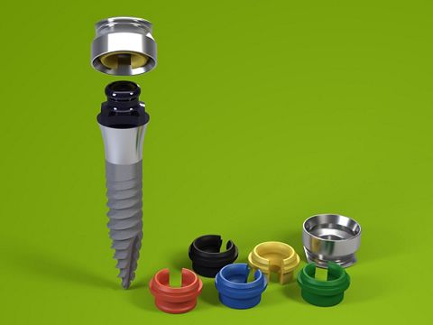
This case shows a good outcome for the management of an edentulous situation, with four Mini Implants inserted in the lower jaw and with early loading. After three months, the control x-ray showed healthy osseointegration of the mini-implants, as well as good gingival healing (photo 10). At this stage the yellow retention inserts (light retention force) were replaced with green retention inserts (medium retention force), giving the patient more confidence to enjoy his meals.
Conclusion
The practitioner was satisfied with the surgical procedure and treatment outcome with the Mini Implants. The surgery was comfortable for the patient as the treatment was finalized fairly quickly, in a single surgery and without bone augmentation. The patient was satisfied with the esthetic outcome and now enjoys a regular diet, which was not possible before due to the poor retention with a conventional denture and soft tissue discomfort. The Mini Implant System has proven to be a good alternative to bone augmentation and will be the preferred option specially for edentulous patients with a difficult anatomic situation.


 . Reducing invasiveness