Treatment of multiple recessions with a modified coronally advanced flap
A clinical case report by Adrian Kasaj, Germany
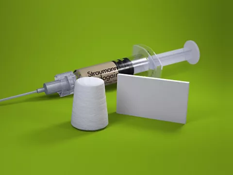
A 29-year-old female patient presented at our clinic with the desire for an esthetic improvement of her teeth. Teeth 1.2 to 1.4 showed multiple Miller class I recessions with a particularly deep recession of >6mm at tooth 1.3 (Fig. 1). The patient was healthy with good oral hygiene. She raised concerns against the harvesting of a connective tissue graft from the palate; therefore, we decided to treat the recessions with botiss mucoderm®, an acellular dermal collagen matrix which is derived from the dermis of pigs through a multi-stage wet-chemical cleaning process. It can be applied instead of an autologous tissue graft in various situations, including the augmentation of attached gingiva and covering of gingival recessions.
Author: Adrian Kasaj

Adrian Kasaj
Dr. med. dent/PhD
Specialist in periodontology (European Dental Association). Associate Professor at the Department of Operative Dentistry and Periodontology, University of Mainz, Germany. Vice Chairman of the “Neue Arbeitsgruppe Parodontologie e.V” (NagP). Author and co-author of more than 80 scientific publications within the field of periodontology. Numerous national and international courses and lectures in the fields of regenerative periodontal therapy and plastic periodontal surgery.

Procedure
The exposed tooth roots were prepared with an air scaler and then treated with 24 % EDTA for 2 minutes. The flap was performed according to Zucchelli with two angular incisions, avoiding the need for vertical incisions (Fig. 2). The anatomical papillae were de-epithelialized. The mucoderm® was rehydrated for 7 minutes in sterile saline solution to enable sufficient flexibility for the adaptation of the matrix to the tooth roots. The matrix was then fixed to the periosteum with cross sutures (Fig. 3). Subsequently, the flap was coronally repositioned and fixed with polypropylene 6-0 sutures (Premilene by B. Braun Melsungen AG). The surgically created papillae were sutured over the de-epithelialized anatomical papillae. Particular attention was paid to complete coverage of the collagen matrix (Fig. 4). Post-operative care included rinsing with 0.12 % CHX solution twice a day and 600 mg ibuprofen if needed. Furthermore, the patient was briefed to avoid tooth brushing in the affected region for 14 days. Sutures were removed 10 days post-operative. The healing time was uneventful. The control at 3 months post-op demonstrated almost complete coverage of the previously exposed tooth roots as well as clear thickening of the marginal gingiva (Fig. 5). In region 1.3 to 1.4, an area of dense connective tissue was visible with a cleft marking the area were the mucoderm® matrix was sutured. Eighteen months post-op, this irregularity has disappeared, without the need for gingival plastic surgery. The gingiva were homogenous and bright (Fig. 6). The patient was very satisfied with the esthetic result.
Discussion
In the last two years, we have treated more than 50 patients with the mucoderm® membrane. Due to very good and predictable results, we prefer applying the matrix using either the tunnel technique or the Zucchelli approach. In any case, good flap mobilization is very important to allow for complete coverage of the matrix and a tension-free closure, both of which are essential for a successful and esthetic outcome. mucoderm® is an acellular matrix which needs to be revitalized. As revitalization from the underlying tooth roots is not possible, an ingrowth of vessels from the covering flap should be ensured. Insufficient mobilization with tensions on the flap could lead to early exposure of the matrix and hence its degradation. An interesting observation that we made in many cases was an improvement in the esthetic outcome progressing for several months after the surgical treatment. A kind of creeping substitution as well as the leveling of irregularities in the thickness of the gingiva without any plastic surgery intervention can be observed.