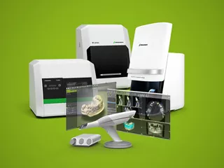
Immediate placement in the esthetic zone followed by a final restoration using Straumann® CARES® Digital Solutions
A clinical case report by André Callegari, Brazil
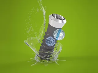
Despite the complications in this case, resulting in the need to activate the implant on the eighteenth day post-placement, the Straumann® Bone Level Roxolid® SLActive® provided a very satisfactory outcome, demonstrating its stability, superior strength and surface treatment, even under extreme circumstances.
Author: André Callegari

Dr. André Callegari
André Callegari is a specialist in dental prosthetics, currently studying for his PhD in Dentistry. He is a founding partner of the Clinic Beleza do Sorriso, in São Paulo, Brazil and coordinator and author of the books Especialidade em foco, vol. 1 and vol. 2 Ed. Napoleão.
Related products and topics


Initial situation
A 21-year-old male patient presented to the Clinic Beleza do Sorriso in São Paulo, Brazil, with a fractured element 21. He was dissatisfied with the esthetics in the anterior segment of the maxilla and had multiple active carious lesions in the interproximal regions of teeth 11, 12 and 22 (Figs. 1,2).
Treatment planning
The aim of planning is to set out a path to ensure the best possible outcome for a specific goal. In high performance dentistry, diagnosis and treatment planning are key elements of success. The first step in this case was to evaluate the patient’s medical history and to confirm that he was in good general health. When starting any esthetic rehabilitation treatment, a number of aspects must be considered: bone architecture, periodontal biotype, interproximal bone crest level and smile line. Plaster study models were then created, which, along with photographic documentation and imaging examinations, helped to identify the root fracture on tooth 21 and slight buccal bone loss. We used the concept DRP (Digital Reverse Planning) which allows full virtual planning, from the bone defect arising from the tooth extraction up to the final outcome of the ideal three-dimensional positioning of the implant in order to obtain the desired esthetic result. We opted for the extraction of tooth 21 and immediate implant placement. All the necessary information obtained through tomography was transferred to the software and a virtual guide was accurately designed. The customized guide was milled without metal washers and based on precise measurements so as to ensure precision placement of the Bone Level Roxolid® SLActive® 3.3 x 14mm implant (Figs. 3-5). After placement, the implant was immediately temporized; the adjacent decayed teeth were properly cleaned and prepared for ceramic fragments in order to correct dental rotations and enhance the esthetics of the anterior region.
Surgical procedure
The planning of the immediate implant enables us to reduce treatment time, leading to greater patient satisfaction and less bone resorption, as well as optimizing function and esthetics from the very first surgical step. In order to achieve successful implant treatments, a review of the literature shows that minimally traumatic surgery is essential, thus preserving the alveolar architecture and surrounding soft tissue.After probing the proximal bone crests, we performed the extraction of tooth 21 and careful socket curettage. The customized surgical guide was adapted and stabilized (Fig. 6); the placement followed the predetermined position and recommended torque for the Bone Level Roxolid® SLActive® implant, selected for the combination of Roxolid® material and the SLActive® surface that increases the resistance for small diameter implants. This provides greater confidence and peace of mind for the resolution of anatomically challenging cases. The occlusal adjustment is a crucial step for a predictable case outcome, to ensure that forces are not transmitted to the implant sooner than expected, thus compromising the final result of the procedure. The patient must receive proper instruction in hygiene and postoperative care (Figs. 7-9).
Unexpected situation
Unexpectedly, 18 days after the implant placement, the patient had a cut-contusion lesion with loss of substance of the lower lip. He reported having undergone direct physical assault with a blunt object. The examination showed considerable mobility of the provisional implant prosthesis, albeit without signs of inflammation in the region. The provisional prosthesis was removed and, to our pleasant surprise, it was found that implant stability had been maintained and only the prosthesis screw had loosened due to the trauma suffered. Immediately, in a single step, we proceeded with the preparation of the ceramic prosthetic parts and the impression of the whole region, including the implant, as described below (Figs. 10,11).
Prosthetic procedure
The excellent treatment surface of the Roxolid® SLActive® implant allows definitive implant activation to be initiated within 21 days. In this case, however, due to the circumstances, the rehabilitation procedures began at 18 days. Ceramic fragments were prepared for teeth 12, 11 and 21 with diamond drills and ultrasonic tips; tissue control was carried out in the region and the impression post was installed on the implant and customized, so that the surrounding soft tissue could be accurately reproduced (Figs. 12,13). The CADCAM system technology allows amazing accuracy, providing better functional and economic benefits to patients. The implant prosthesis was made following the CARES® approach on the Straumann® Variobase® abutment, which allows exact engagement of the coping to the Straumann® Variobase® abutment with its four engaging grooves. For scanning and modelling of the case, the Dental Wings platform was used to design the implant prosthesis and the ceramic for the adjacent teeth (Figs. 14,15). The milling was done in zirconia for later application of ceramic coverage, characterization and customization of the elements. The parts were tested in the mouth and, after adjustment, were properly etched and cemented (Figs. 16-21).
Final result
4 months’ follow-up; stable surrounding tissues and occlusion; satisfied patient and satisfactory radiographic appearance (Figs. 22-25).