Collagen membranes in regenerative dental medicine
A scientific essay by Sascha Böhm and Sandra Wrobel, botiss biomaterials
Guided tissue and guided bone regeneration (GTR, GBR) are well-established techniques in dentistry to augment lost tissue around teeth and dental implants respectively (Nyman et al. 1980, Karing et al. 1980, Nyman et al. 1982, Dahlin et al. 1988). The basic principle of these methods is the placement of a barrier membrane between the soft tissue and residual bone in order to prevent the fast-proliferating epithelial cells from populating the bony defect and to provide space and time for the migration of slow-dividing osteogenic or periodontal ligament cells into the defect area.
Authors: Sascha Böhm & Sandra Wrobel
Dr. Sascha Böhm and Dr. Sandra Wrobel are Scientific Product Managers at botiss biomaterials, Berlin/Germany.
Related products
Introduction
In the course of the evolution of GTR and GBR techniques, different types of membranes have been developed. Today, commercially available barrier membranes for GTR or GBR procedures can be divided into either non-resorbable or resorbable membranes. Non-resorbable membranes, which were first introduced in the dental field, are primarily made of titanium and polytetrafluoroethylene (PTFE, expanded PTFE and dense PTFE) and maintain their structural integrity over the entire healing period. Despite their successful use in many clinical indications, primary soft tissue closure for certain defects remained challenging and additional surgery to remove the device after the desired healing time was inevitably required (Zucchelli and Mounssif 2000, Wang et al. 2000). On the contrary, bioabsorbable membranes, either of synthetic (aliphatic polyesters) or natural (collagen) origin, are metabolized by hydrolysis or enzymatic activity respectively, and thus are completely resorbed over time, increasing patient comfort.
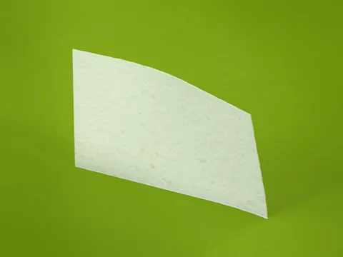
Jason® membrane and collprotect® mebrane
botiss Jason® membrane and botiss collprotect® membrane – two collagen membranes available in the Straumann Biomaterials portfolio – are made of porcine collagen type I and III. Both membranes are produced in a standardized multi-stage cleaning process that removes all cellular and non-collagenic components, while preserving the native three-dimensional and open porous collagen structure. In this regard, tolerance studies as well as clinical trials have proven their biocompatibility (Rothamel et al. 2012, Barbeck et al. 2015, Merli et al. 2015, Panagiotou et al. 2015).
Requirements, type and characteristics of a barrier membrane
Practically speaking, a barrier membrane designed for dental indications should meet the following criteria: host tissue integration, biocompatibility, cell occlusiveness, permeability for nutrients and ease of use (Hardwick et al. 1994). Since collagen is a major and highly conserved protein found in the connective tissues of all mammals, thus offering a high level of homology, different collagen membranes for GTR or GBR procedures of animal origin have been engineered. From 28 different collagen types that have been identified in vertebrates, collagen type I is the most prevalent and best-described member of the entire family (Shoulders and Raines 2009, Fratzl 2008, Kadler et al. 2007). In humans, collagens cover ~30% of the absolute protein content of the body and can be found in all tissues and organs such as skin, bone, tendon, etc. They are important proteins involved in numerous biological activities including extracellular matrix- and blood vessel formation, cell adhesion and migration, as well as tissue morphogenesis and repair (Kadler et al. 2007, Sakar et al. 2012). They are present as elongated fibers, network-forming collagens, and fibril-associated collagens or as transmembraneous collagen domains (Shoulders and Raines 2010, Fratzl 2008) and give stability and elasticity to the tissue by their remarkable tensile strength. Collagen molecules are synthetized by different cells, such as endothelial (Howard et al. 1976) or smooth muscle cells (Schlumberger et al. 1991), but predominantly by fibroblasts (Silvipriya et al. 2015).
Degradation of collagen and its function in tissue remodeling and wound healing
Specific proteases (collagenases) regulate the cleavage of collagen molecules. With their characteristic catalytic domains they are able to decompose the molecule into defined fragments. Degradation and biosynthesis of collagen are essential steps for several processes. Unlike other substances or compounds used for tissue engineering approaches, natural collagens are very effective biomaterials due to their fast adaptation to mechanical forces and the transformation of information into biomechanical signals, thereby controlling several events e.g. tissue remodeling or wound healing (Chang and Buehler 2014). Collagens also play a major role in hemostatic reactions. Platelets harbor cell surface-exposed collagen-specific receptors and binding of a ligand induces degranulation and blood clotting. Hence, collagen aids in wound stabilization and for this reason has been widely used as a hemostatic agent and biological dressing in medicine and pharmacology (Patino et al. 2002, Nuyttens et al. 2011). In addition, it acts as chemoattractant for different cell types involved in the wound healing process, including gingival and periodontal ligament fibroblasts (Postlethwaite et al. 1978). When exposed to the oral cavity during healing time, collagen is rapidly degraded in a non-inflammatory manner, making the use of collagen membranes possible even in challenging flap procedures (Schwarz et al. 2006).
Collagen membranes for GBR/GTR in dental medicine
Collagen membranes used in dental medicine derive mostly from tendon, dermis, skin or the pericardium and are commonly of bovine or porcine origin (Bunyaratavej et al 2001). They can be manufactured by different techniques. In general, the collagen fraction is first isolated, then purified and precipitated by changing the ionic strength, the pH value or by increasing the temperature followed by an air evaporation step. At the end, the purified collagen is freeze-dried and sterilized (Patino 2002).
Mechanical behavior and degradation time offer unparalleled range of treatment options
Even though they derive from the same animal species, Jason® membrane and collprotect® membrane differ in their mechanical properties and resorption behavior, since they originate from different porcine tissues. Jason® membrane derives from the porcine pericardium, a fibrous tissue sac that surrounds the mammalian heart. The pericardium prevents overexpansion of the heart and contains small amounts of serous liquid, enabling free mobility of the heart muscle during blood volume changes. Since the pericardium has to withstand the forces of the heart muscle, it has an exceptionally dense collagenic structure, which confers rigidity and a multi-directional tear resistance as well as tensile strength to the membrane. Thus, in bone augmentation procedures, Jason® membrane undergoes slow enzymatic degradation and consequently provides an extended barrier time, making this membrane suitable for the treatment of larger defects, such as extended ridge augmentations and sinus floor elevation with additional lateral augmentation. Extended bony defects require a prolonged healing time as the complete resolution of the defect is dependent on the rate of blood vessel formation and recruitment of bone-forming cells, which starts at the edges and proceeds to the center of the defect area (Schenk et al. 1992). Conclusively, the dimension of the defect ultimately determines the time a membrane is desired to maintain its barrier function (Zellin et al. 1995). Accordingly, smaller and medium-sized defects such as fenestrations or periodontal lesions require a membrane with intermediate barrier time, provided by the collprotect® membrane showing a faster degradation compared to Jason® membrane. The collprotect® membrane is made of porcine dermis and has an open porous, but also a dense collagen network. The inherent open pores of the native porcine skin facilitate the migration of blood vessels into the defect area, hence allowing a rapid vascularization of the underlying wound bed (Rothamel et al. 2011), while the density of the membrane maintains a barrier against soft tissue ingrowth. Aside from their different performances as mechanical barriers, Jason® membrane and collprotect® membrane further differ in their thickness. Jason® membrane has a low thickness of about 0.2 mm and facilitates soft tissue manipulation especially in thin biotypes. collprotect® membrane is slightly thicker (~0.4 mm) and may be the material of choice for augmentation with autologous or allogenic bone. These kind of grafts usually completely remodel in 3 to 4 months and therefore do not require a prolonged barrier function. Similarly, barrier membranes with intermediate barrier times, such as collprotect® membrane, are also adequate for the treatment of periodontal bone defects.
Periodontal regeneration following treatment with barrier membranes
Resorbable collagen membranes have been successfully used to treat periodontal intrabony lesions and furcation defects. Compared to conventional flap surgery, an average gain of clinical attachment (CAL) of 1.1 mm to 1.58 mm was achieved when a collagen barrier membrane was placed between the gingival epithelium and the residual intact periodontal ligament (Needleman et al. 2002, Needleman et al. 2006, Stoecklin-Wasmer et al. 2013). Moreover, systematic reviews indicate similar clinical outcomes compared to non-resorbable membranes in regenerative periodontal therapy (Laurell et al. 1998, Parrish et al. 2009), however without the drawbacks of non-resorbable material. Because of their stiffness, non-resorbable membranes are prone to flap perforation and increase the risk of soft tissue dehiscence and membrane exposure, impairing the regenerative process and ultimately requiring the removal of the device (Simion et al. 1994, Gher et al. 1994, Watzinger et al. 2000).
Convenient membrane solutions due to excellent handling properties
In addition to their mechanical and degradation behavior, the Jason® and collprotect® membrane also feature excellent handling and defect applications. Both materials can be applied wet or dry. In dry conditions, the relative stiffness of the membranes allows upright placement into the defect or socket during filling with particulate bone graft material. Accordingly, after rehydration with sterile saline solution or blood, they become flexible and can readily be placed over the augmentation area and adapted to the surface contours. If needed, repositioning in wet conditions is easily possible without the risk of the membrane sticking to itself. Although both membranes can be pinned, a fixation is unnecessary in most cases due to their excellent adhesion properties. Owing to its exceptionel tear resistance, Jason® membrane can even be sutured or screwed. Likewise, both membranes can easily be trimmed with scissors or a scalpel to fit the defect area. Modern techniques in dental medicine aim at both optimal tissue regeneration and satisfaction of esthetic concerns. Over the past decades, barrier membranes made of collagen have been demonstrated to be central to achieving these goals. Both the Jason® membrane and collprotect® membrane used in more than 300,000 dental applications exhibit outstanding handling properties, have controlled degradation patterns and are characterized by their excellent biocompatibility, making these membranes the ideal choice for applications in implant dentistry and periodontology.