“I have always aimed to carry out anterior esthetic corrections as a preliminary stage before the actual prosthetic treatment,” explains Swantje Matthes, dental technician and dentist, and adds: “I don’t work on cross-bites or prepare major dental realignment. I prefer to refer the patient to an orthodontic specialist or other specialist.” The team of doctors at the owner-managed joint dental practice in Cologne-Nipps where Matthes has been working for some six years now covers the entire range of dentistry. “I see myself as an all-rounder with prosthetic experience.”
At the start of 2019, she realized that ClearCorrect was the ideal supplementary treatment option for her prosthetic patients. “Of course, patients benefit if I can preserve more tooth substance in the preparation, by resolving crowding and shaping the dental arches, and even out the ceramic layer thickness of the restoration, so that the overall outcome is more esthetic.” It is also “a benefit, that I can offer my patients a five-year warranty, if they do happen to need follow-up treatment, so they don’t need to pay any extra.” The Unlimited option from ClearCorrect covers both the aligner and the retainer fixed rate for five years.
ClearCorrect aligners are made of a fracture-resistant material (0.76 mm polyurethane) with high retention properties that is resistant to discoloration. As the material is transparent, the aligners are discreet and almost invisible, which means they are very much favored by esthetically conscious patients. Eating habits are not restricted either, as the aligners can be removed. This also means patients do not need to make any adjustments to their dental hygiene routines. The aligners also feature a smooth, straight trimline that, unlike other aligners, extends beyond the margins of the gingiva. The higher retention strengths this achieves means that fewer attachments are required1. The tongue quickly gets used to the aligner, so patients soon sound perfectly normally to both wearers themselves and listeners. Matthes: “I have tried it out myself and found it to be successful.” The treatment period depends on the extent of correction and the period the aligner is worn (22 hours a day, but at least 19 hours), and varies from one person to another from between four and 24 months. “For my prosthetic patients, I allow for a preliminary treatment of up to six months.” After consulting the treatment provider, the aligners are generally changed every 14 days. You can see how successful the treatment is even while it is still ongoing, and the teeth move gradually into the desired position. “DenToGo is a smartphone app that makes it easier to monitor and follow-up aligner therapy remotely,” explains Matthes. “The system detects when the next correction step is due based on photographs that the patients take themselves.”

Want to stay up to date?
youTooth.com is THE PLACE TO BE IN DENTISTRY – subscribe now and receive our monthly newsletter on top hot topics from the world of modern dentistry.
Case study: patient-friendly with digital impressions
Matthes’s first case involved an esthetic anterior correction before prosthetic treatment. The dentist explains: “The 62-year-old patient wanted a new treatment with ceramic crowns in her upper incisors for 12, 11 and 21 and 22. The teeth had been endodontically treated at a different practice about a year and a half ago following trauma to the front teeth and had been reconstructed with plastic, which had already partially splintered away.
The clinical findings in both the upper and lower jaws revealed crowding, with vestibular displacement of tooth 21 and moderate overlapping of the lower incisors. The prosthetic baseline was not ideal. To achieve the best esthetic outcome possible, and as the patient has high esthetic demands, I recommended an orthodontic pre-prosthetic therapy with clear aligners, to create enough space for the preparation so it can hold the crown. The patient consented that she should have the orthodontic treatment before the prosthetic restoration, for which we factored in about six months, and in August 2019 X-rays and photographs were taken (anterior and profile views and the occlusal view of the maxilla and mandible). The situation scans of the upper and lower jaws were also taken with the 3Shape Trios Intraoral scanner.”
The conventional method of taking impressions of the upper and lower jaws can, in essence, be used to produce the aligners; however, ClearCorrect offers the option of a digital workflow, which is “both user-friendly and patient-friendly,” explains Matthes. “The modern alternative to conventional impression taking is also much more comfortable for patients; there is no gagging or unpleasant taste, with shorter treatment times, as the impression can be taken with precision and speed.” When it comes to prosthetic treatments, adds Matthes, who now uses digital impressions for almost all her prosthetic patient cases (“I only take a conventional approach for removable prosthetics”), “it improves communication between the patient, the dentist and the dental technician.” At this point, she emphasizes that “you need a decent lab that knows its way around digital workflows. Distance is no obstacle. Our dental lab, “Echt Eppers” is in Hildesheim, more than 300 kilometers (185 miles) away – but everything still runs perfectly and it also means there is no chain of infection; a valuable asset in these times of corona.”
Even if aligner creation is not about imaging the preparation margins: “Even here, for simple situation scans, where you are mapping the dental situation, it is really important to make sure the mirror and lenses are clean” - practical advice from the dentist who has been working with Trios since 2017. “Mirrors that don’t reflect anything after they are sterilized so many times, are - to put it simply - a waste of time.” The photo and scan data are then uploaded as an STL file via the 3Shape portal, from where the state-of-the-art production site for the ClearCorrect aligners in Texas*, USA, can then open the scans. “Then I enter information in the computer on the selected treatment methods, including information on whether an approximal enamel reduction is planned, or if the use of engagers, attachments or aids for specific tooth movements, are approved,” she explains.
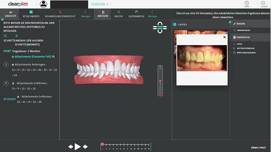
Then the data are analyzed and a treatment simulation is created. “I looked at it with the patient, and she was able to follow the phases on the monitor and see what tooth movements were planned for the next six months.” This preview can also be forwarded directly to the patient as a link. Then the customized ClearCorrect aligners are manufactured specifically for the patient based on the treatment plan. Matthes: “The treatment plan consisted of eight aligners for the upper and lower jaws. In week 4, five attachments or engagers were added in both the upper and lower teeth to aid treatment.” There were check-ups every four to six weeks. After around six months the treatment was completed. “The patient wore the last pair of aligners as retainers at night.” As the front teeth in the mandible exhibited grade I mobility, we decided to wait with the next step. A few weeks after the preliminary orthodontic therapy, there was no movement in the lower anterior teeth, and so the preparation of the upper teeth could be terminated.

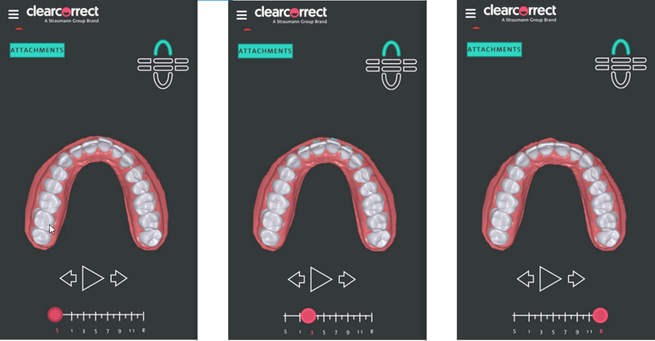
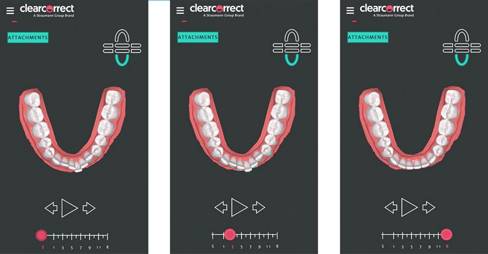
Fig. 3: 3D simulation for the patient Occlusal view of the maxilla and mandible. Before treatment is initiated and the aligner is produced, ClearCorrect sends a treatment plan (3D simulation) covering each step of aligner treatment that allows a targeted doctor-patient consent discussion. Here the ClearCorrect treatment plan is based on eight aligners (worn for 14 days each). The computer preview on the left shows the baseline. In check-up week 4, engagers (attachments) are recommended (vestibular green marking on the teeth).

Fig. 3: 3D simulation for the patient Occlusal view of the maxilla and mandible. Before treatment is initiated and the aligner is produced, ClearCorrect sends a treatment plan (3D simulation) covering each step of aligner treatment that allows a targeted doctor-patient consent discussion. Here the ClearCorrect treatment plan is based on eight aligners (worn for 14 days each). The computer preview on the left shows the baseline. In check-up week 4, engagers (attachments) are recommended (vestibular green marking on the teeth).
*Aligner production started in Germany at the production site in Markkleeberg near Leipzig in January 2021.
Conclusions for clinical practice
Aligner therapy with ClearCorrect is a valuable addition for general dentists to offer in their practice and can be used in a very patient-oriented approach, for instance to correct minor misalignment or open an existing gap before prosthetic treatment. “It is easy to learn, making it suitable for anyone interested in the technique,” is how Matthes sums up her experience with ClearCorrect. Online courses with experienced aligner users can make it even easier to launch into the technique, as the speakers not only provide basic, practice-based orthodontic training but also pass on their experience with ClearCorrect2,3. The option of a digital workflow with digital impression-taking (intraoral scan), data transfer and remote monitoring is a benefit for the dentist and the patient and supports a contemporary treatment concept. “Overall, this procedure lays the foundation for a minimally invasive prosthetic approach,” Matthes sums up, “which also paves the way for a successful, overall esthetic outcome.”
For on-demand professional development and webinars see www.clear-correct.de/education

