


What is this about?
- How precise implant placement in the maxillary molar region can be achieved using the Straumann Falcon™ dynamic navigation system combined with the Straumann® Fast Molar Solution.
- Step-by-step integration of digital data acquisition, prosthetically driven planning, and dynamically navigated osteotomy preparation to ensure accuracy and predictability.
- How a fully digital and navigated workflow contributes to efficient treatment, reduced surgical risk, and reliable functional outcomes in implant rehabilitation.
Introduction
Posterior maxillary tooth loss is one of the most frequent clinical indications in implant dentistry. Patients increasingly expect an efficient solution that combines short treatment times with a high level of safety and predictability. This expectation is particularly relevant in the molar region, where functional demands are high and delayed rehabilitation can negatively affect mastication and overall quality of life. From a clinical perspective, posterior sites often present anatomical challenges such as limited vertical bone height, proximity to the maxillary sinus, and reduced bone quality, which increase surgical complexity and the risk of deviations from the planned implant position.
In this context, the treatment requires more than accelerated treatment protocols. It relies on precise diagnosis, prosthetically driven digital planning, and accurate surgical execution to ensure predictable outcomes. The integration of digital workflows enables clinicians to manage complex posterior indications efficiently, even in patients with systemic conditions, while maintaining a minimally invasive approach and a high standard of care.
The Straumann Falcon™ dynamic navigation system represents an important advancement in achieving these objectives. By providing real-time intraoperative guidance on osteotomy preparation, including angulation and drilling depth, the system supports a high level of surgical accuracy while preserving intraoperative flexibility. When combined with comprehensive digital planning and integrated services such as Smile in a Box®, the Straumann Falcon™ dynamic navigation system allows for optimized osteotomy preparation in the posterior maxilla and a seamless transition between surgical and prosthetic steps.
The use of the Straumann® Anatomic Healing Abutment (AHA) further enhances this digital workflow by supporting guided soft-tissue healing immediately after implant placement. Designed to follow the natural emergence profile of the tooth, the AHA promotes stable peri-implant soft-tissue contours, reduces the need for additional soft-tissue manipulation, and minimizes chairside adjustments during the prosthetic phase. This contributes to improved esthetic integration, enhanced patient comfort, and a more efficient overall treatment process.
The following case report shows the application of a fully digital workflow using the Straumann Falcon™ dynamic navigation system in combination with the Anatomic Healing Abutment to deliver a precise, predictable, and efficient Straumann® Fast Molar Solution in the posterior maxilla. The case highlights how digital planning, dynamic navigation, immediate postsurgical data acquisition, and optimized soft-tissue management can be combined to achieve stable functional and esthetic outcomes, even in a medically compromised patient.
Initial situation
A 55-year-old female patient presented to our clinic. The patient’s chief complaint was the rehabilitation of the edentulous space resulting from the extraction of tooth #26, with the expectation of a fast and safe treatment approach leading to favorable functional and satisfactory esthetic outcomes.
The medical history revealed von Willebrand disease (VWD), chronic asthma, arterial hypertension, and a documented allergy. Current medication included candesartan (Atacand®, Germany) for the management of high blood pressure. The patient reported no history of smoking.
Regarding dental history, tooth #26 was extracted on 29 April 2024 due to a vertical root fracture. In addition, the patient had previously undergone implant placement in the region of tooth #17, followed by successful prosthetic rehabilitation with an implant-supported crown between 2022 and 2023.
The current clinical examination showed a plaque index of 27%, with no bleeding on probing, no suppuration, and no other clinical signs of inflammation. Overall oral hygiene and patient compliance were considered good. It was observed that tooth #26 was absent. The clinical examination indicated that the edentulous area likely had sufficient mesiodistal and buccopalatal space for implant placement (Figs. 1,2).
A radiographic assessment, including a panoramic x-ray and cone-beam computed tomography (CBCT), together with digital treatment planning, demonstrated a vertical bone height of approximately 6 mm, indicating the need for an internal sinus floor elevation, and a horizontal bone width of 13.2 mm. The bone quality was classified as D3 (Fig. 3).
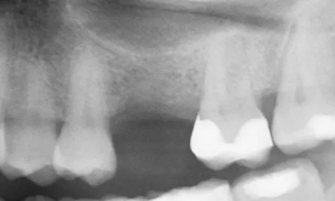
Fig. 3
The prognosis for the remaining teeth was considered good based on both clinical and radiographic findings, supported by adequate oral hygiene and good patient compliance. According to the ITI SAC classification, the case was categorized as having medium surgical complexity and complex prosthodontic complexity (Fig. 4).
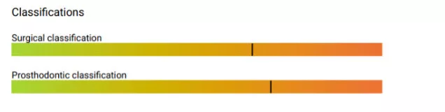
Fig. 4
Treatment planning
- The Straumann® Smile in a Box® service was used for prosthetically driven digital planning and fabrication of the Straumann Falcon™ surgical marker or surgical guide?
A comprehensive digital treatment plan was established. An intraoral scan (IOS) dataset was obtained, and the point cloud data were preserved for further use (Fig. 5).

Fig. 5
A CBCT scan of the maxilla (field of view 80 × 50 mm) was created to allow three-dimensional visualization of the bony structures. The IOS and CBCT datasets were imported into the coDiagnostiX® software and superimposed in order to perform a prosthetically guided planning of the implant position and the selection of the suitable Straumann® Anatomic Healing Abutment XC (AHA) (Figs. 6-8).
The treatment was planned for dynamically navigated osteotomy preparation using the Straumann Falcon™ dynamic navigation system. In addition to the implant position and AHA selection, the position of the navigation marker was planned, and a corresponding fixation marker was fabricated by means of 3D printing (Rapid Shape). The planning and preparation of the navigated surgery were performed by the Smile in a Box® service (Straumann®) and subsequently verified by our team in the clinic.
The IOS dataset was duplicated and prepared for intraoperative scanning.
- Implant placement with Straumann BLX™ Ø 4.5 x 8.0 mm SLActive®, Roxolid®.
- Placement of Straumann® Anatomic Healing Abutment XC (AHA) RB/WB, Shape XL, Ø 4.5 mm, GH 1.5 mm, H 4.5 mm.
- Directly postsurgical digital scanning performed for the fabrication of the final restoration.
Placement of final restoration.
Surgical procedure
Due to the patient’s von Willebrand disease (VWD), desmopressin (DDAVP, Ferring GmbH, Germany) was administered prior to surgery. In addition, the patient received antibiotic prophylaxis with 1.5 g of amoxicillin (Amoxicillin-ratiopharm®, Ratiopharm GmbH, Germany) one hour before the procedure.
Local anesthesia was achieved using 3.4 ml of articaine hydrochloride /epinephrine hydrochloride at a concentration of 1:200,000 (Ultracain D‑S®, Septodont GmbH, Germany).
The Straumann Falcon™ dynamic navigation system was used to support dynamically navigated osteotomy preparation.
Before starting the surgery, a case check was performed with Straumann Falcon™.

Fig. 9
The surgical approach consisted of a minimally invasive flap design, with a single central crestal incision in the region between teeth 25 and 27.
After raising a small flap, the marker was fixed to the teeth and the implant site preparation was initiated under dynamic navigation (Fig. 9).
After drilling down to the maxillary sinus floor, an internal sinus floor elevation of approximately 2 mm was performed using the Summers technique with a concave osteotome (Ustomed GmbH, Germany) (Fig. 10).

Fig. 10
Subsequently, a Straumann BLX™ implant Ø 4.5 x 8.0 mm, SLActive®, Roxolid®, was placed in the region of tooth #26, following the manufacturer’s instructions. Implant insertion was performed using a contra-angle handpiece, and fine-adjusted manually by ratchet (Figs. 11,12).
After implant placement, a Straumann® Anatomic Healing Abutment XC (AHA) RB/WB Shape XL, ∅ 4.5 mm, GH 1.5 mm, was inserted (Fig. 13).

Fig. 13
Soft tissue closure was achieved with two sutures, one mesial and one distal, using PTFE Coreflon 5/0 sutures (Implacore, Poland) (Fig. 14).

Fig. 14
Following implant placement and suturing, the anatomic healing abutment was scanned intraoperatively and the data were integrated into the original digital planning dataset (Figs. 15,16).
This workflow allowed the fabrication process of the final restoration to begin immediately after surgery.
The AHA screw access channel was then closed with Clip F (Voco GmbH, Germany), and a control radiograph was taken, which confirmed that everything was in the proper position (Fig. 17).
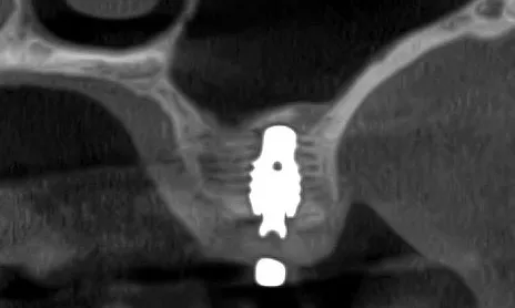
Fig. 17
Postoperatively, desmopressin (DDAVP, Ferring GmbH, Germany) was administered due to the patient’s von Willebrand disease (VWD), and Cyklokapron® mouth rinses (Pfizer Pharma GmbH, Germany) were prescribed to minimize the risk of bleeding. Wound healing was uneventful, and the patient was scheduled for routine follow-up to monitor soft tissue recovery and implant integration.
Prosthetic procedure
The implant was restored following a conventional loading protocol, with the final prosthesis delivered five months after surgery due to the patient's interim stay at another hospital. At the five-month visit, the soft tissue was healthy and ready for the final restoration (Figs. 18,19).
The laboratory fabricated the final crown based on the scans, taken at the day of surgery. The final abutment used was a Straumann® Variobase®, and the restoration was fabricated from milled zirconia (Figs. 20,21).
Due to the predefined anatomical shape of the Straumann® Anatomic Healing Abutment XC, predictable soft-tissue healing was achieved. The emergence profile of the final crown was subsequently designed and fabricated accordingly, allowing placement of the definitive restoration without exerting pressure on the peri-implant soft tissues. Occlusion was carefully adjusted to ensure proper function. A control radiograph was taken, showing satisfactory positioning. (Figs. 22-25).
At the six-month follow-up, healing remained uneventful, and the implant and prosthesis demonstrated stable integration and function.
Treatment outcomes
The treatment was completed successfully without complications, despite the patient’s underlying von Willebrand disease. The patient reported that the procedure was unproblematic and pain-free, with the number of appointments minimized. The use of digital tools within a prosthetically driven workflow, including dynamic navigation, enabled precise and minimally invasive implant placement, while the anatomic healing abutment supported predictable soft-tissue healing. This resulted in a successful prosthetic outcome. From the clinician’s perspective, the outcome was highly satisfactory in terms of esthetics, function, and overall oral health.
Author’s testimonial
“The treatment, from data acquisition to digital planning and navigated osteotomy preparation, was very targeted and completely free of problems. Thanks to the intraoperative scan after insertion of the AHA, the final restoration could already be placed in the next treatment session, which led to a reduction to three appointments for the entire treatment. This enabled a significant increase in efficiency in the digital workflow.”
Kai Vietor
Take-aways
- Prosthetically driven digital planning enhanced predictability in this posterior maxillary case, allowing implant placement to be aligned with the planned restoration despite limited vertical bone height and proximity to the sinus.
- Dynamic navigation with the Straumann Falcon™ system supported accurate, minimally invasive osteotomy preparation, facilitating controlled execution of the planned implant position and sinus floor elevation while reducing surgical trauma.
- The use of a Straumann BLX™ implant with SLActive® surface and Roxolid® material supported reliable clinical performance in a medically compromised patient, providing confidence in early healing and mechanical stability under demanding conditions.
- Soft-tissue management with the Straumann® Anatomic Healing Abutment XC, combined with a Variobase® as the final abutment, enabled a predictable prosthetic workflow, resulting in stable peri-implant tissues and a pressure-free final restoration.
- The workflow described, which utilizes an Anatomic Healing Abutment, has significantly increased efficiency by reducing the number of treatment appointments.