#Full-Arch 22. Nov 2022

Bimaxillary full-arch rehabilitation with Straumann® PURE Ceramic Monotype Implants in an irradiated patient: a 5-year follow-up case report

A clinical case report by Inge De Latte, Ghent, Belgium

Ceramic dental implants are now widely used in implant dentistry. Single, multiple, or full dental arches can be restored with ceramic implants, and this is possible nowadays thanks to the development of surfaces like ZLA®, novel materials, and enhanced treatment protocols.

Introduction

Clinical usage of ceramic dental implants is a reliable treatment alternative to titanium implants, supported by increased scientific evidence. Ceramic dental implants meet the growing desire for metal-free treatments and a natural, highly esthetic look, and address the needs of patients with metal hypersensitivities.

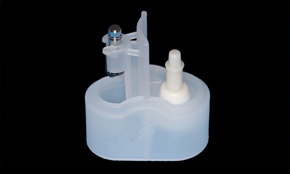
The Straumann® PURE Ceramic Monotype Implant consists of an implant and abutment made from zirconium dioxide ceramic. It is based on features of the Straumann® Soft Tissue Level Standard Plus and Straumann® Bone Level Implants. Therefore, they are very straightforward to implement in our practice, as the surgical cassette used is the same as that for the Straumann® Bone Level Implants.

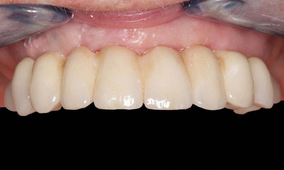
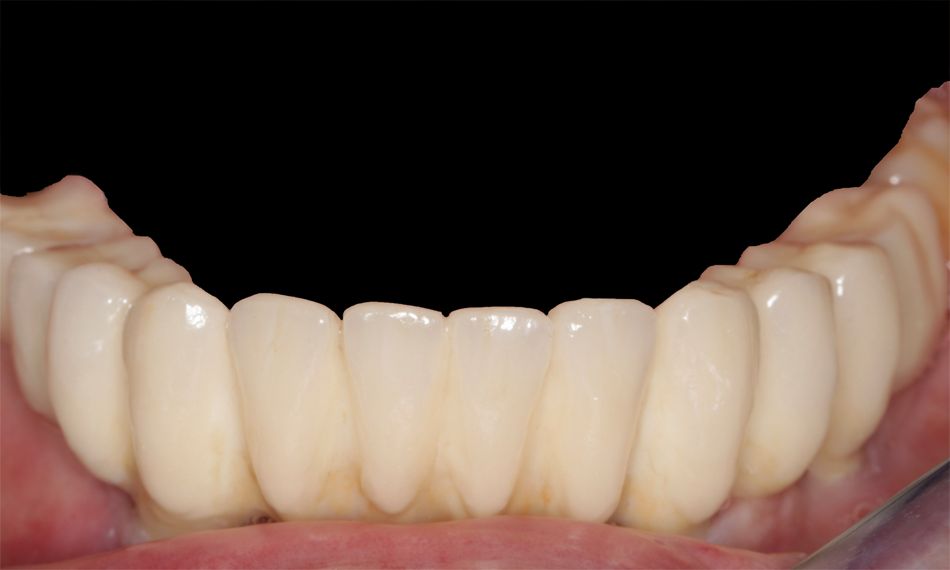
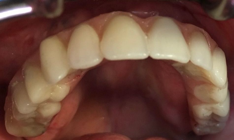
The following clinical case shows a successful bimaxillary full-arch rehabilitation with zirconium bridges in the lower and upper jaws supported, respectively, by four and six Straumann® PURE Ceramic Implants Monotype in a patient who requested a metal-free solution for her unstable prostheses. The patient later received chemotherapy for a tonsil carcinoma. Even 5 years after completing the implant treatment, the patient remained satisfied with the outcome and did not report any mechanical or biological complications during the follow-up visits.

Initial situation

A 70-year-old female patient presented herself to our clinic in 2014. Her main complaints concerned unstable lower and upper restorations that did not allow her to chew or speak properly; moreover, she explained that these issues also affected her general quality of life and self-confidence. Her medical history revealed alopecia due to an underlying autoimmune disorder. In addition, she reported poly-allergic features to nickel and emphasized her desire to keep a fixed bridge with a metal-free solution for her peace of mind.

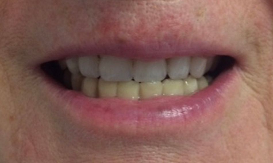
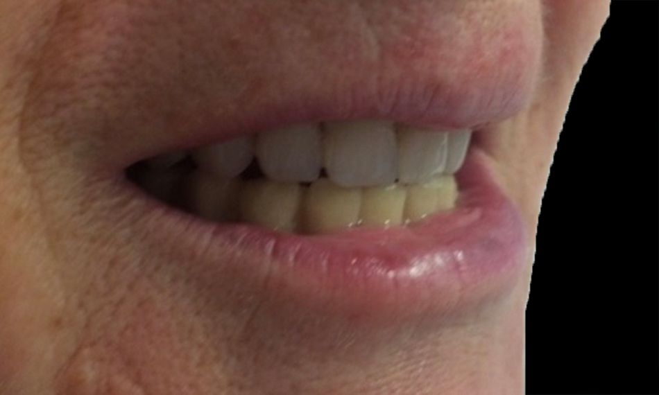
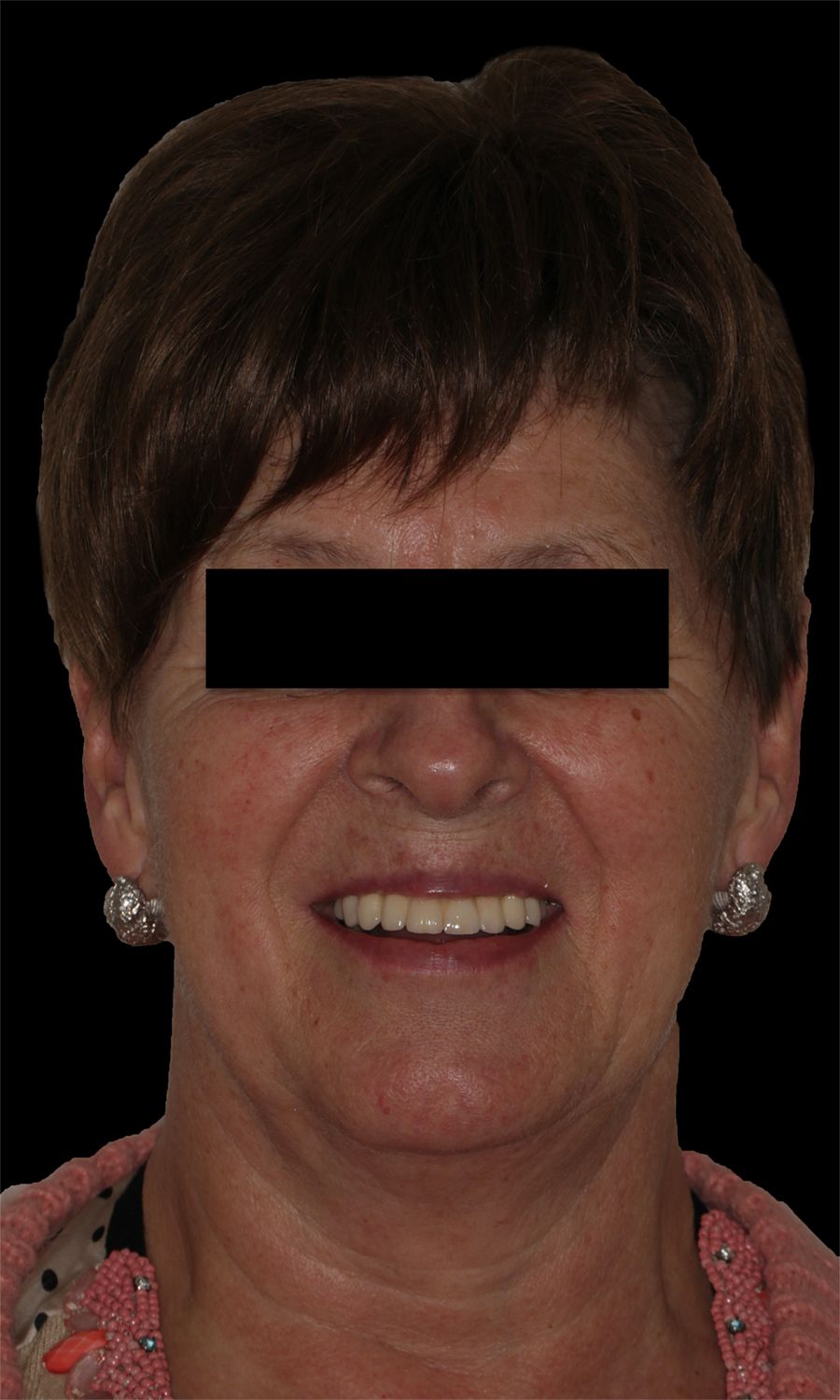
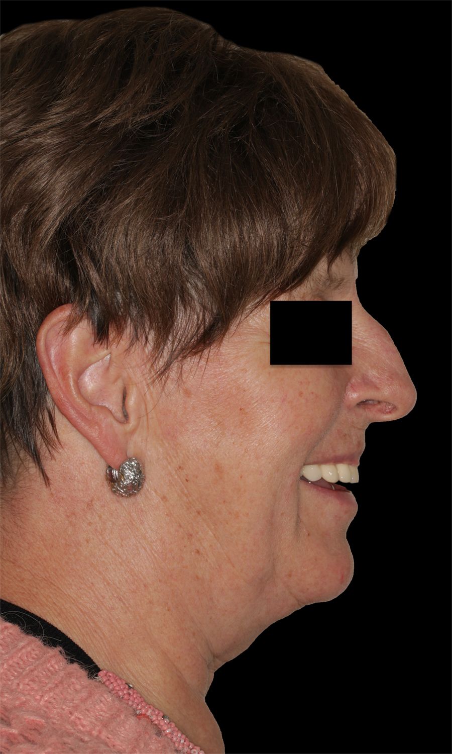
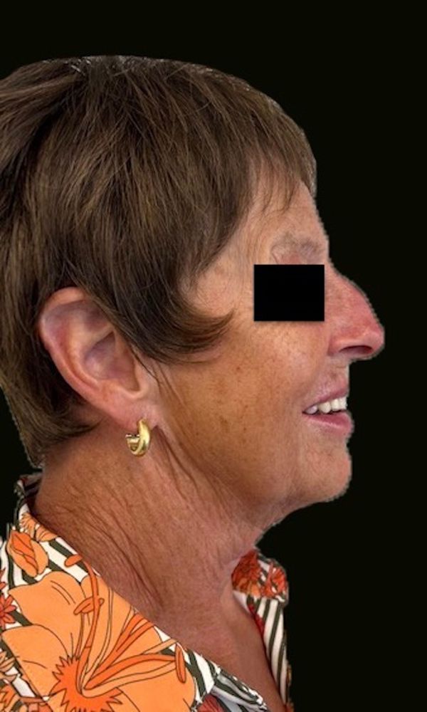
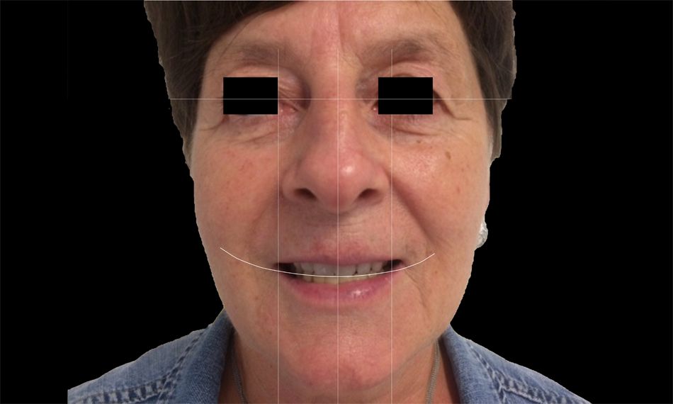
The extraoral examination showed mild face asymmetry and an average smile line (Figs. 1-3).

The intraoral examination showed an edge-to-edge bite with upper midline deviation. The patient was periodontally compromised on the remaining teeth, with the presence of deep periodontal pockets, inflammation, dental mobility, and bleeding on probing on all sites, except on teeth #37 and #47.
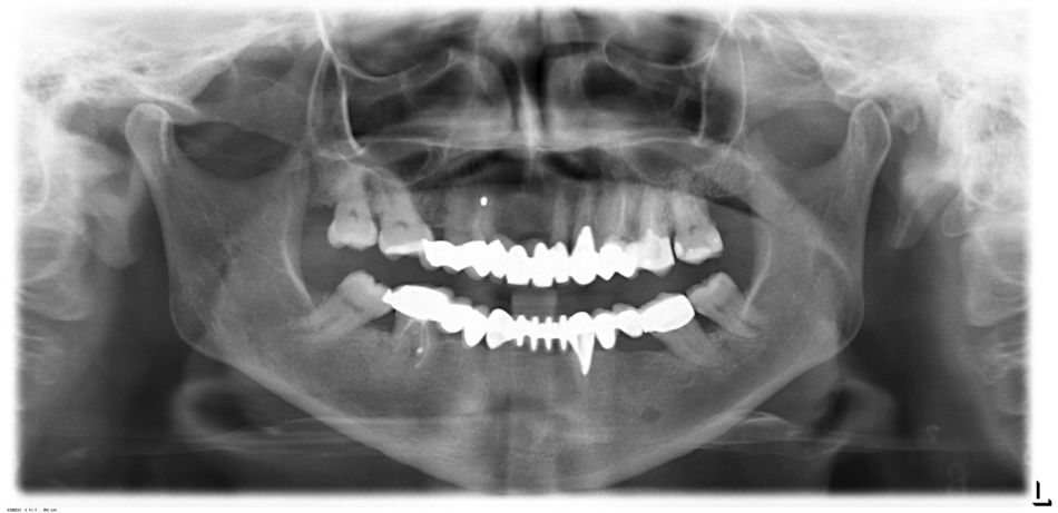
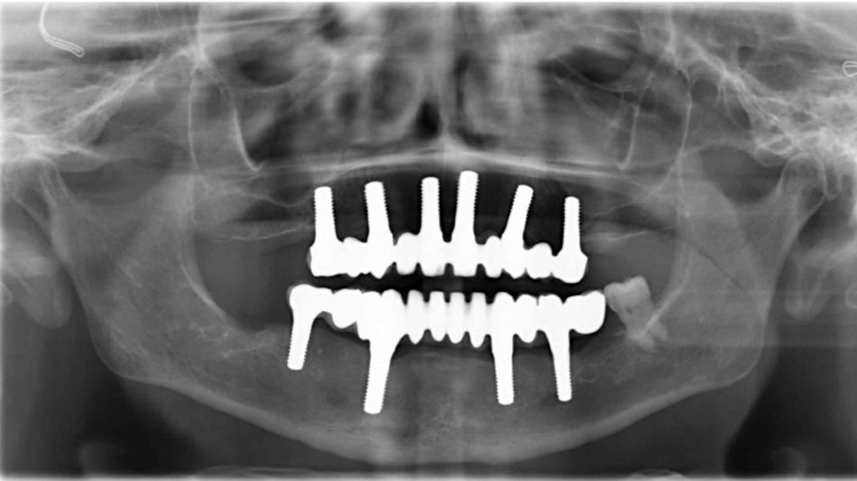
The radiographic assessment revealed bridge structures with generalized bone loss and apical infections in some locations (Fig. 4).

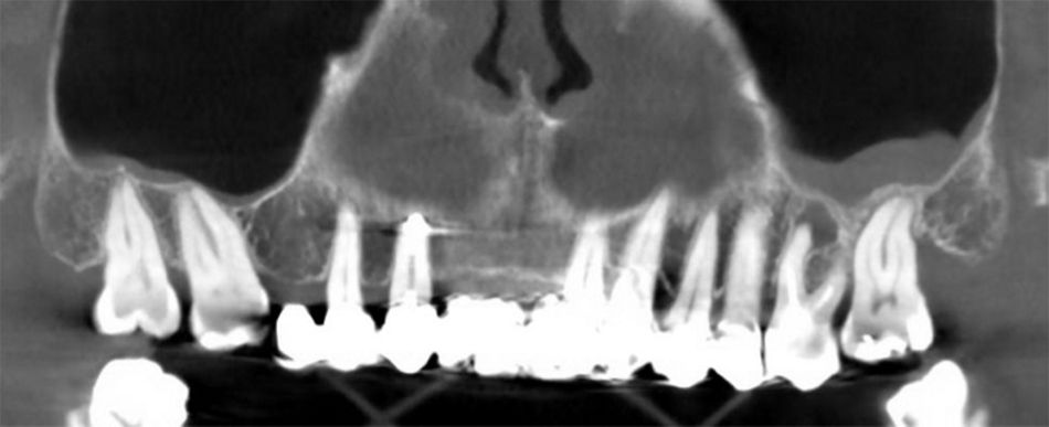
Following our clinical protocol, a tomogram was taken to properly confirm a diagnosis and for digital case planning (Fig. 5).

Treatment planning

After a thorough discussion of the various treatment options, and taking into consideration the requirements and medical conditions of the patient and her desire for a metal-free solution, zirconium structures were chosen instead of titanium. We collectively decided to place a full zirconium bridge, with six Straumann® PURE Ceramic Monotype Implants in the upper jaw and four in the lower jaw, extracting the compromised teeth and maintaining the periodontally stable teeth #37 and #47.
Due to the active infection and discomfort for the patient, the non-viable teeth in the lower jaw were extracted in the first phase. The procedure was done under local anesthesia, consisting of 2% lidocaine and 1:100,000 epinephrine. The conservative removal of the roots was the main goal of the atraumatic dental extractions, as we aimed to maintain as much buccal bone height as possible, while preserving alveolar crestal height in all dimensions. Teeth #37 and #47 were maintained and, as requested by the patient, an immediate provisional prosthesis was provided for the healing phase until the day of implant placement.
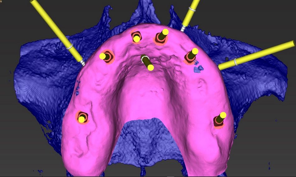
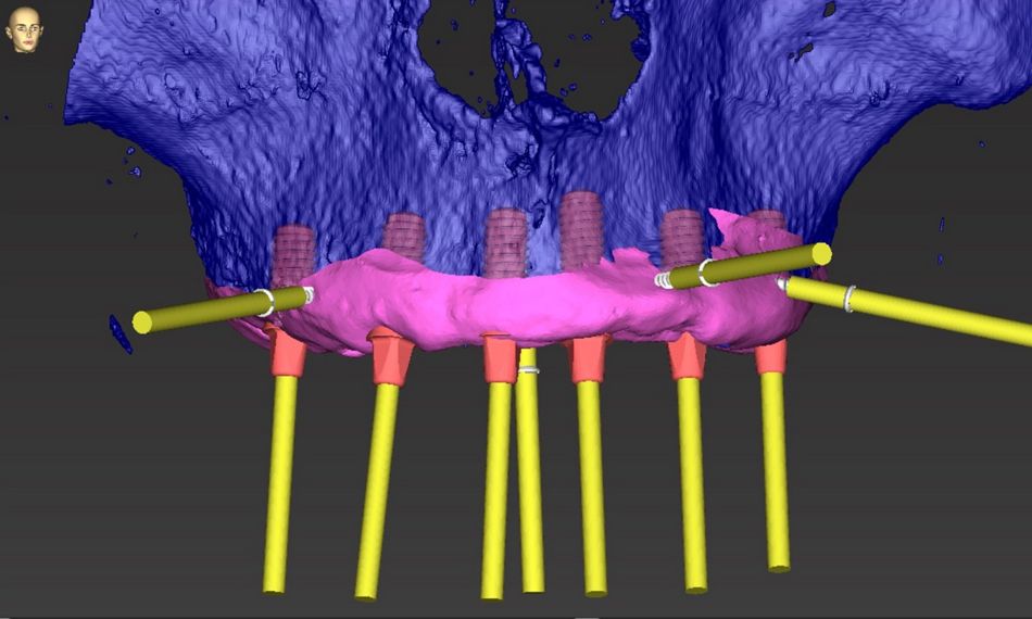
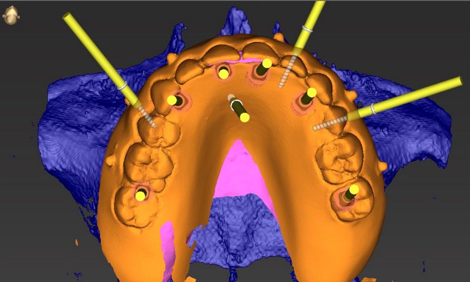
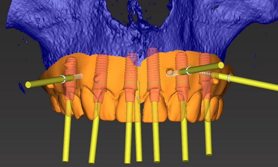
Impressions were taken and, with the aid of coDiagnostiX®, implants were digitally planned in an optimal prosthetic-driven position (Figs. 6-9). The surgical guides were printed with high-resolution fluid for surgical guides and prepared with sleeves and pins by the dental lab.

Surgical procedure

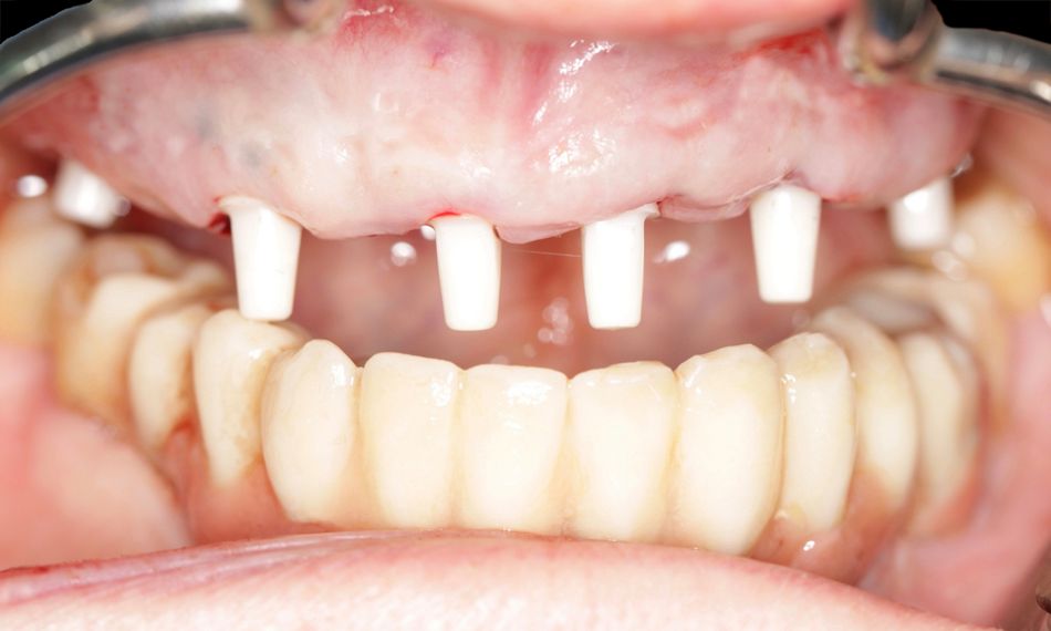
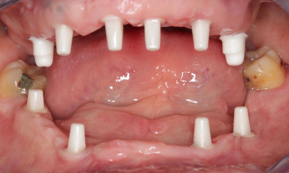
After the healing period of two months, four Straumann® PURE Ceramic Monotype Implants were placed in the lower jaw, in positions #34, #36, #43, and #46. We used Straumann® PURE Ceramic Monotype Implants with an abutment height (AH) of 5.5 mm (diameter 4.1 mm – ZLA, 10 mm in positions # 34, #36, and #46, 12mm in position #43). The temporary prefabricated bridge was adapted and placed chairside using the RD PURE Ceramic Temporary Coping – PMMA - for crowns.

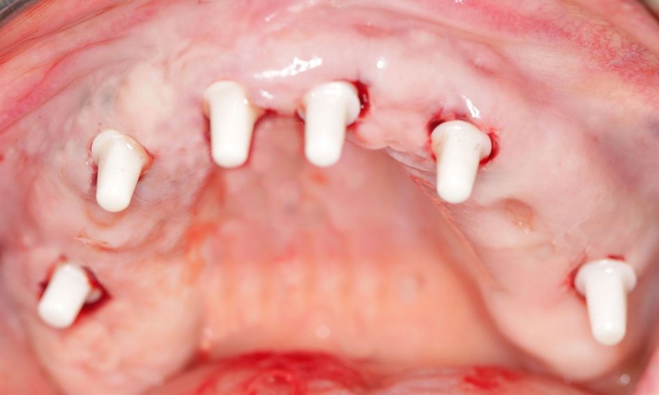
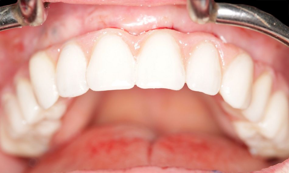
Likewise, in the upper jaw, non-salvageable teeth were extracted six months later in a conservative manner. Two months after extraction, six Straumann® PURE Ceramic Monotype Implants were placed in positions #16, #13, #11, #21, #23, and #26. We used Straumann® PURE Ceramic Monotype Implants with an AH of 4 mm, except for #26, where we used an AH of 5.5 mm. The implant diameter was 4.1 mm, and the implant length was 10 mm in positions #16, #13, #11, #23, and #26, and 12 mm in position #21.

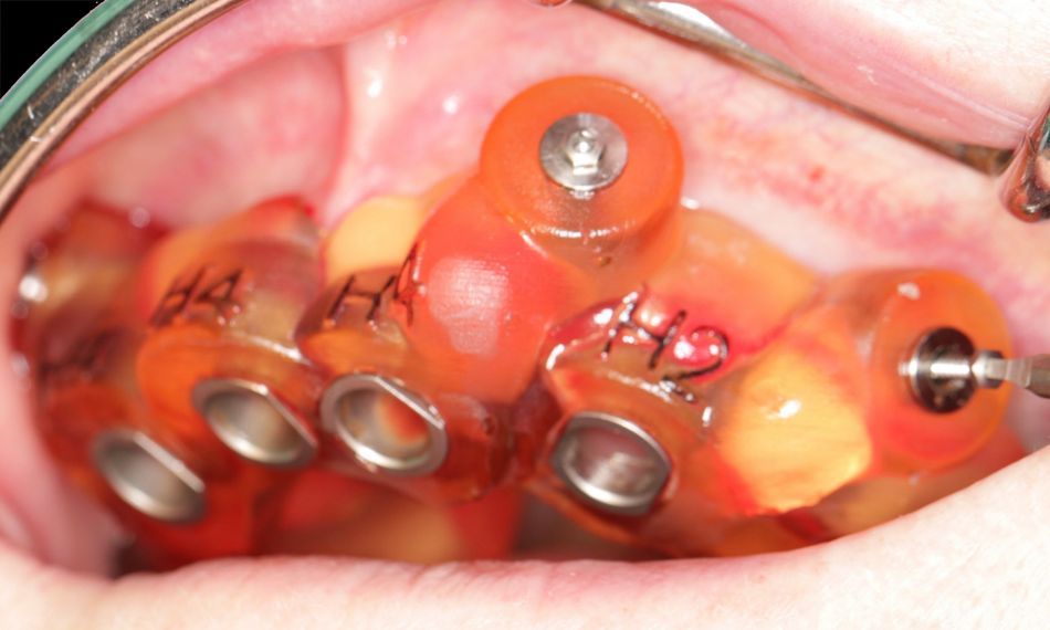
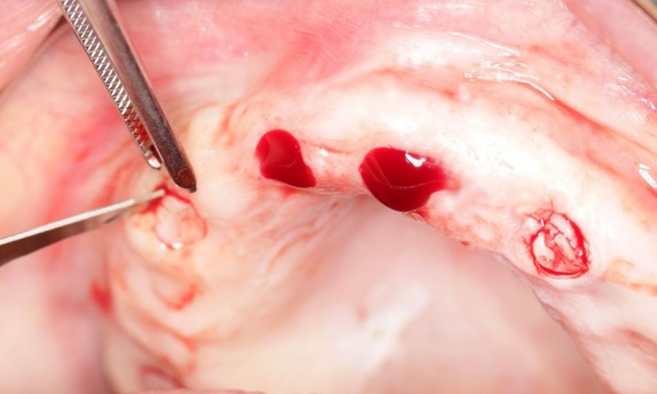
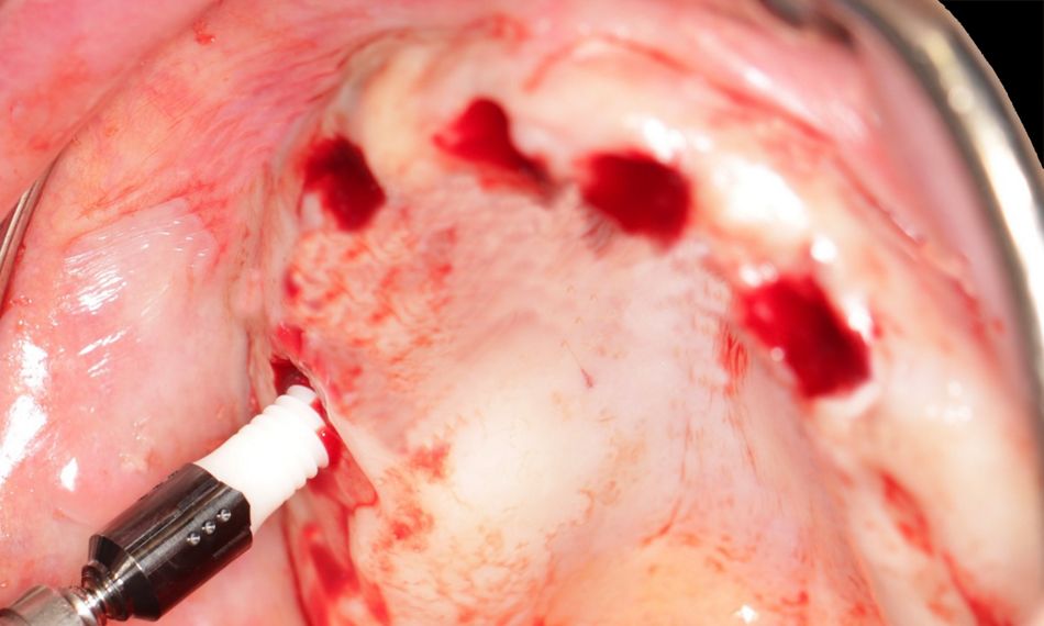
On the day of surgery, the surgical guide was placed, and its optimal fit was checked. Local anesthesia, consisting of 2% lidocaine and 1:100,000 epinephrine, was infiltrated. After the guide’s fixation pin was drilled and inserted, the mucosa punch was used to remove the soft tissue conservatively (Figs. 10-12).

For the implant bed preparation, the manufacturer’s instructions were followed according to the protocol provided by coDiagnostiX®. The surgical protocol for osteotomy preparation for Straumann® PURE Ceramic Monotype Implant is the same as for the corresponding bone level implant, as the implant body mimics the Straumann® Bone Level design.

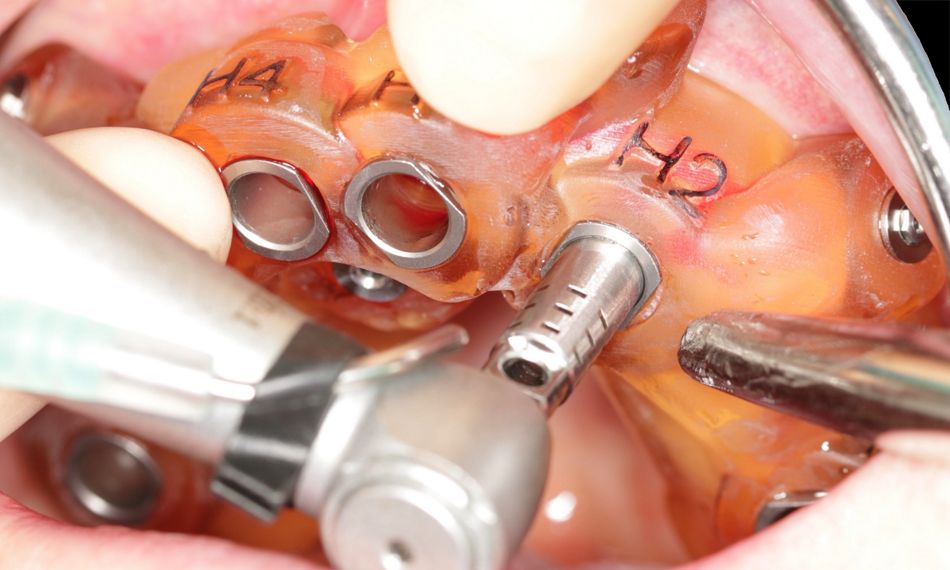
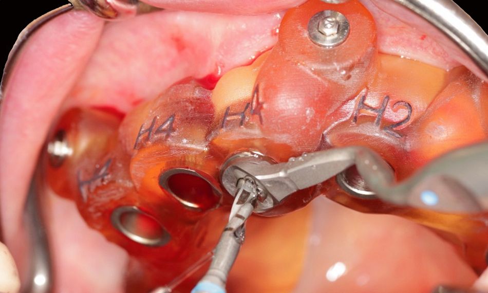
Pilot and twist drills were used to prepare the implant bed in its basic form: the final drill size for the 3.3 mm implant was 2.8 mm, and the final drill size for the 4.1 mm implant was 3.5 mm. Utilizing the appropriate profile drills and taps for the 3.3 mm and 4.1 mm diameter implants, the implant bed was finally prepared (Fig. 13).

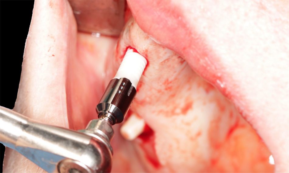
Following the implant bed preparation, the guides were removed, and the implants were placed freehand. The PURE monotype implant comes with a separate transfer piece that locks securely into place, and this fact also contributed to their precise placement in the planned prosthetically driven position (Figs. 14-18).

Following implant placement, and as requested by the patient, immediate provisional restorations were provided, and the occlusion was checked (Fig. 19). A temporary prefabricated bridge was adapted and placed chairside using the RD PURE Ceramic Temporary Coping – PMMA. The provisional PMMA bridges click onto the ceramic implants. No temporary cement was needed.

Prosthetic procedure

Before starting the final prosthesis, we evaluated the temporary ones. The patient’s wish was for the anterior teeth to be more visible, and this request was considered for the final restoration.

Impressions were taken with the closed impression technique.

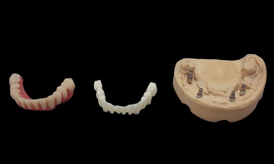
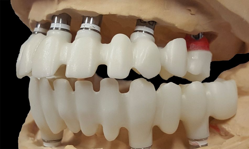
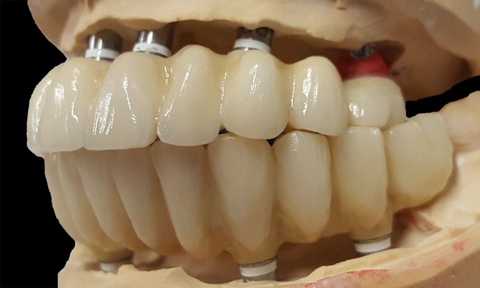
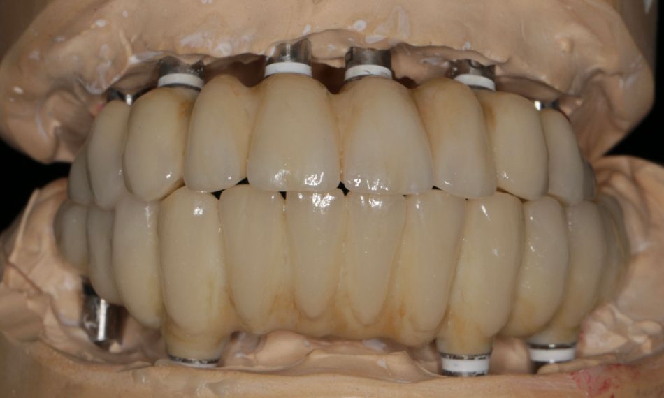
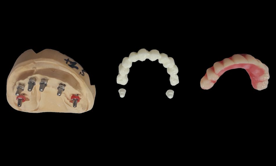
The impressions were sent to the laboratory, and implant analogs were fixed and models made. The models were placed in an articulator with temporary bridges on the models. In the next step, a try-in was done, with more visible teeth than the temporary ones (Figs. 20-21).

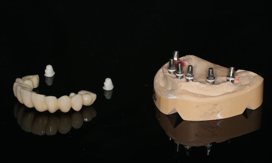
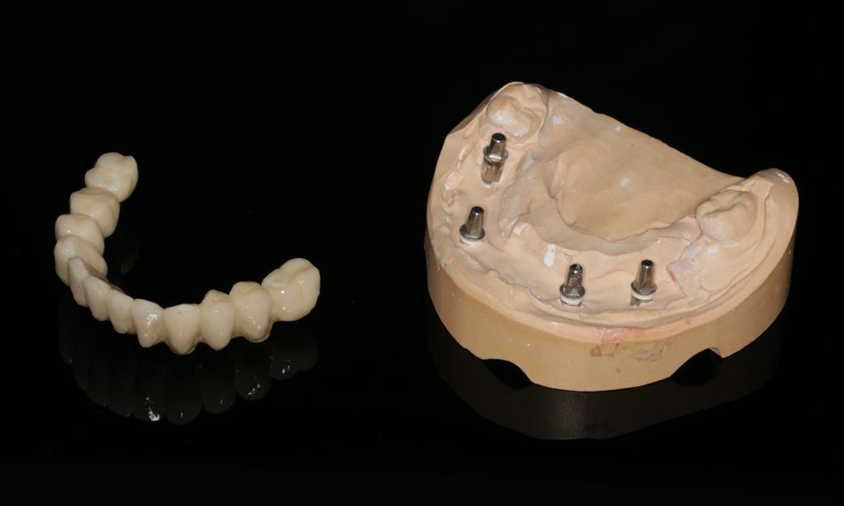
The patient and dentist liked the esthetic result. On that basis, the lab fabricated the zirconium dioxide structures in one piece for each arch. The zirconium structures were designed using the Straumann Dental Wings software. The models and try-in were scanned, and the margin lines were defined. Finally, the teeth were placed, and the form was adjusted.

The milling was performed using a Straumann® Zirconia blank in a Straumann® CARES® M Series by Amann Girrbach milling system. On these structures, porcelain VM 9 from Vita was used (Figs. 22-26).

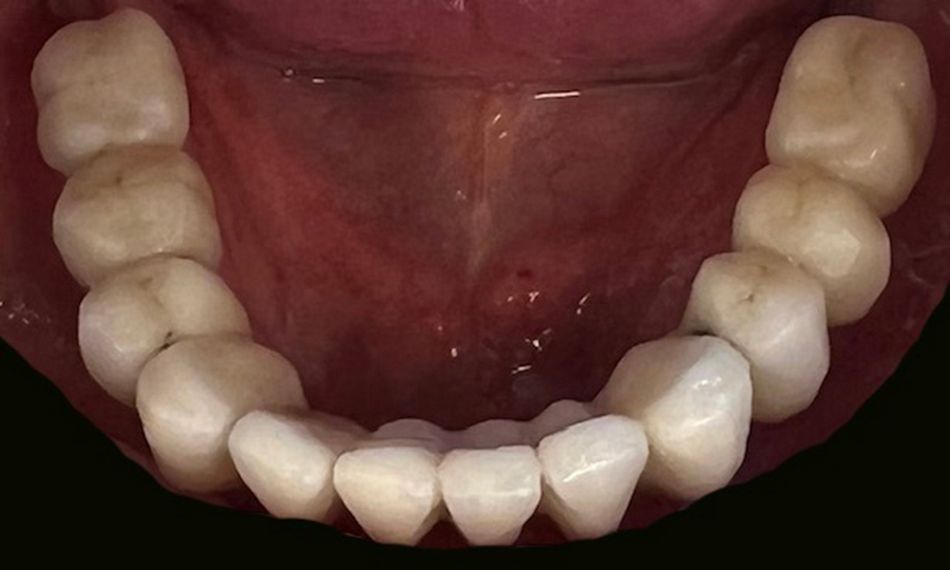
The patient came to the clinic for the placement of the final restoration. During this visit, the healing of the tissues was evaluated, and the prosthesis was tried. The abutment part of the Straumann® PURE Ceramic Implants was cleaned and prepared for cementation. After cementation, all excess cement was removed, and oral hygiene instructions were provided (Figs. 27-29).

Follow-up visits were arranged one week and one month after cementation. No further adjustments were needed, and no biological or mechanical complications were reported (Figs. 30-32).


Want to stay up to date?

youTooth.com is THE PLACE TO BE IN DENTISTRY – subscribe now and receive our monthly newsletter on top hot topics from the world of modern dentistry.


Treatment outcomes

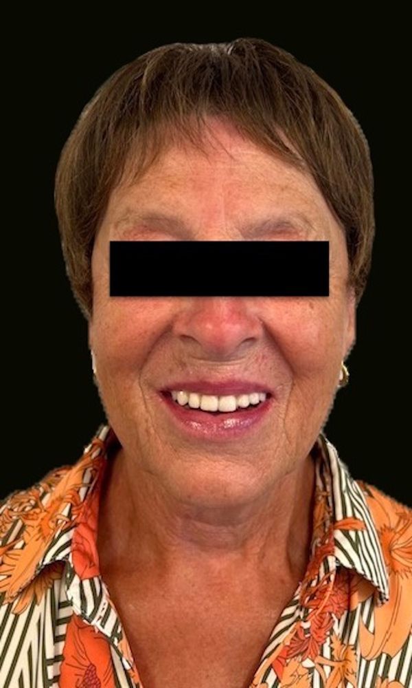
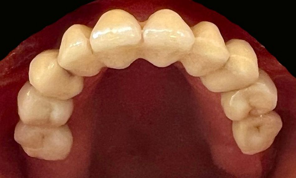
One year after implant placement, a spinocellular carcinoma of the throat was diagnosed. The patient was treated with 70 Gy radiation. The patient developed a change of taste and a dry mouth, but the implants remained stable. After five years, the patient is very satisfied with the treatment outcome regarding the esthetics, function, and health. In addition, she did not report any biological or mechanical complications and is involved in a follow-up and maintenance program every three months that involves clinical and radiographic (yearly) assessments and reinforcement of oral hygiene instructions (Figs. 33-37).

Author's testimonial

Although this was a very challenging case, I enjoyed every moment. Considering the fact that the patient was treated with radiotherapy for throat cancer one year after implant placement, the PURE Zirconium implant proved to be the most stable and effective implant I have ever seen during my whole career. I am happy that we had the opportunity to do such a beautiful, high-level treatment. The follow-up during 5 years of a medically compromised oral situation, in particular after a challenging radiotherapy, made me realize that we need more research and more future cases to prove the biologically ultra-friendly, long-term stability of the Zirconium PURE implants in compromised patients.

I sincerely want to thank the prosthetic dentist, Jean-Pierre Cornelis, and the dental lab LABO HOET GHENT and the head of the lab, Mieke Van Ooteghem, for the support, time, patience, and technical skills to complete this case.


A Center of Dental Education (CoDE) is part of a group of independent dental centers all over the world that offer excellence in oral healthcare by providing the most advanced treatment procedures based on the best available literature and the latest technology. CoDEs are where science meets practice in a real-world clinical environment.