
Introduction
The rise and spread of digital dentistry have significantly influenced clinicians who specialize in implant dentistry and full-arch restorations.3 The continued development of CAD/CAM technology and 3D printing has revolutionized the manufacture of tooth-borne and implant-supported fixed dental prostheses.4
Digital technologies for fabricating fixed full dental prostheses with implants can result in less expensive laboratory and clinical chairside time and lower overall cost.5
Furthermore, due to its excellent survival rate, immediately loaded implants with a fixed prosthesis have become a feasible choice in the rehabilitation of edentulous patients.6
The following case report aims to show a successful treatment with immediate loading of a final fixed restoration in the edentulous maxilla with Straumann® BLX implants.
Initial situation
A systemically healthy, non-smoking 59-year-old female with no medication or allergies attended our clinic with the following chief complaint: “I have been wearing full dentures for many years, but now I find that they come loose easily, which prevents me from eating and speaking normally. Would it be possible to improve my situation without going through complex surgeries or waiting a long time?“
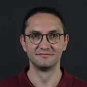
The extraoral examination with the denture depicted a medium smile line and loss of the occluding vertical dimension due to the wear of the denture. Without the denture, inadequate soft tissue support, which affected the facial esthetics, was apparent (Fig. 1).
The intraoral examination showed that the supporting soft tissues were slightly inflamed. The alveolar ridge of the maxilla and mandible showed a loss in width and height, although it was still sufficiently wide and covered with relatively healthy keratinized mucosa (Fig. 2).
The initial radiographic evaluation included an orthopantomogram (Fig. 3) and a cone-beam computed tomography scan (CBCT). The CBCT scan was used to determine the amount and density of bone for implant placement in the edentulous areas. Sagittal cuts revealed sufficient bone availability for implant placement, although the bone was of medium quality (type 3).
The SAC classification was used to determine the degree of difficulty of the implant-related rehabilitation for the patient. The patient was classified as a complex case from the surgical and prosthodontic standpoints (Fig. 4).
Treatment planning
Following the clinical and radiographic examination, and considering the patient's desire and health, it was decided to place six Straumann® BLX implants through the mucosa-supported guide by the flapless surgical procedure and to perform an implant-supported prosthesis.
The treatment workflow included:
1. Prosthetic and esthetic analysis.
2. Digital planning with coDiagnostiX® (Fig. 5) and mucosa-supported guide design and preparation (Fig. 6).
3. Placement of six Straumann® BLX implants through the mucosa-supported guide.
4. Prosthesis prototype in resin-based provisional material made with the Straumann® 3D printer and with attached Variobase® copings.
5. Immediate loading of Straumann® BLX implants.
6. Final monolithic zirconia screw-retained prosthesis preparation.
A Center of Dental Education (CoDE) is part of a group of independent dental centers all over the world that offer excellence in oral healthcare by providing the most advanced treatment procedures based on the best available literature and the latest technology. CoDEs are where science meets practice in a real-world clinical environment.
Surgical procedure
The mucosa-supported guide was checked on the edentulous area for proper fit. The flapless surgery was performed under local anesthesia with 2% lidocaine and 1:100,000 epinephrine. The fixation pins were then drilled and inserted (Fig. 7). The flapless technique was used for implant placement. The implant bed was prepared using the Straumann® Surgical Cassette following the pilot drilling protocol (Fig. 8). The implants were placed manually with the ratchet in a clockwise direction with a final torque of 35-50 Ncm (Fig. 9). Position indicators on the implant driver and surgical guide were aligned as anticipated to indicate implant depth and connection orientation as planned in coDiagnostix® (Fig. 10).
Following the implant bed preparation, six flapless guided Straumann® BLX implants were inserted (Fig. 11). Primary stability was achieved in all implant sites, which allowed us to proceed to the planned immediate loading protocol.
Prosthetic procedure
17-degree angled Straumann® Screw-Retained Abutments (SRA) with a rounded shape and designed for an emergence profile were placed on the implants in positions #13, #14, and #23. In addition, straight SRAs were placed on the implants in positions #16, #24, and #26 (Fig. 12). The Straumann® SRA connection ensures long-term mechanical stability and precision against rotation. The SRAs are intended to allow versatility, especially when treating edentulous patients with Straumann® BLX implants.
Open-tray impression copings were splinted together using Teflon and light-cured pattern resin (Fig. 13). During the laboratory phase, protective caps were placed to protect the implants and preserve the soft tissue shape (Fig. 14).
Next, the lab technician fabricated the prosthesis prototype (Fig. 15).
The prosthesis prototype was produced from resin-based provisional material with the Straumann® 3D printer (Fig. 16), which increased the efficiency and shortened the workflow. Variobase® copings were attached to the prototype.
Two days after the surgery, the prosthesis prototype was screwed onto the abutments. Passive fit, occlusion, vertical dimension, position of the teeth, and the relationships between the teeth and soft tissues were evaluated intraorally and extraorally (Figs. 17, 18).
The prosthesis was produced monolithically using Straumann® yttria(Y2O3)-stabilized tetragonal zirconia polycrystal (Y-TZP) blocks from the prototype STL file prepared for the design of the prosthesis prototype via CAD-CAM technology (Fig. 19). After sintering and glazing procedures, Variobase® copings were cemented to the hybrid prosthesis with self-adhesive resin cement. The gingival part of the hybrid prosthesis was stained using Optiglaze™ (Fig. 20).
The patient was followed up six days after surgery. The healing was uneventful (Fig. 21), and the hybrid prosthesis was screwed onto the abutments in the patient’s mouth (Fig. 22).
The proper extension and the prescribed occlusal scheme were checked. A mutually protected occlusion with anterior guidance was achieved (Figs. 23,24).
Eight months after implant placement, the patient returned for a follow-up visit (Fig. 25), and an orthopantomogram was taken (Fig. 26). Furthermore, the patient was involved in a yearly maintenance program, where oral hygiene instructions were reinforced, and a complete clinical and radiographic assessment was performed.
Treatment outcomes
This clinical case report describes the successful management and outstanding outcome of an edentulous patient who desired a fixed, fast, and atraumatic solution. The patient received six Straumann® BLX implants and was treated with the immediate loading of the final fixed restoration. The treatment was completed in 6 days. The patient did not have to take any medication as the morbidity was excellent. Finally, the facial esthetics was improved due to the adequate soft tissue support.

Want to stay up to date?
youTooth.com is THE PLACE TO BE IN DENTISTRY – subscribe now and receive our monthly newsletter on top hot topics from the world of modern dentistry.
Author’s testimonial
Taking advantage of the digital workflow, dental implants can be placed with a flapless approach without disturbing the soft tissue integrity. As a result, the final prosthesis can be loaded immediately, and the total treatment time is significantly shorter. In this case, the process of placing six Straumann® BLX implants with an immediate fixed prosthesis delivery was completed in only six days.