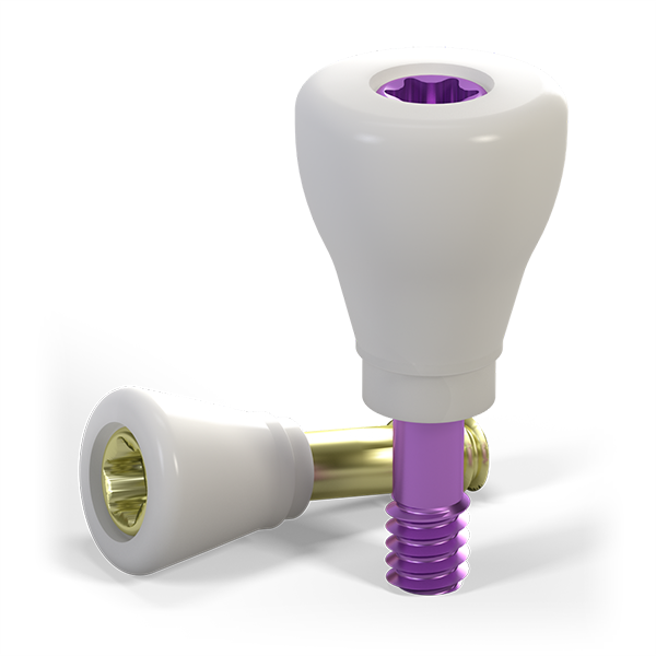
Healing abutments are a unique type of abutment designed to facilitate the healing of bone and soft tissue around a dental implant. Once the gingiva has healed (generally 4-6 weeks after implant placement), the healing abutment is replaced with the final abutment that connects the implant to the restoration.
Abutment material
A wealth of research has shown that the abutment material itself has a significant role to play in peri-implant soft tissue health.1Healing abutments are currently available in a wide range of materials; the most common pre-manufactured stock healing abutments are made of titanium, ceramic and PMMA. Zirconia (zirconium dioxide, ZrO2) has been recognized as an exceptional material for implant abutments with mechanical properties comparable to the “gold standard” titanium.2

Want to stay up to date?
youTooth.com is THE PLACE TO BE IN DENTISTRY – subscribe now and receive our monthly newsletter on top hot topics from the world of modern dentistry.
Clinical advantages
Ceramic healing abutments are comparable to natural teeth and superior to titanium abutments and exhibit a range of clinical advantages.3 These include enhanced blood flow in tissues surrounding zirconia abutments (an indicator for the health of the soft tissue around implants), which translates into better nutrition of the tissues surrounding the implant.
The long-term survival of dental implants depends, among the other factors, on the control of bacterial infiltration in the peri-implant region.4 It has been observed that ceramic surfaces exhibit lower bacterial colonization on the implant-abutment interface5, 6 and a statistically significant decrease in human plaque biofilm formation compared to titanium surfaces.7 This reduces nitric oxide synthesis (an indicator of the bacteria-induced inflammatory process) in tissues around zirconia healing abutments.8
A systematic review that evaluated the impact of the abutment characteristics on peri-implant tissue health discovered significantly less bleeding on probing (BOP) and plaque accumulation with zirconia when compared to titanium abutments.1 Moreover, a comparison of the quality of the soft tissue surrounding zirconia and titanium abutments demonstrated lower inflammatory activity around the former5, 6. This finding was later confirmed by a pre-clinical study, which reported a lower proportion of pro-inflammatory leucocytes in the epithelium around zirconia than titanium abutments, indicating a superior gingival seal.9 Finally, zirconia surfaces also exhibited a faster formation of the epithelial attachments in vitro and a higher degree of soft tissue integration than titanium.10
Conclusion
In conclusion, the abutment material type plays a significant role in soft tissue healing and maintenance. As a highly soft-tissue-friendly material, zirconia could be considered a material of choice when successful soft tissue healing is essential.