#Esthetics 28. Apr 2023

Bone augmentation procedures and implant placement in the esthetic zone

A clinical case report by Dr. Juli Pan, China

Immediate implants have emerged as a solution that offers numerous benefits to patients, including shorter treatment times and less morbidity; however, this approach is not always possible or indicated.

Introduction

Tooth extraction and long-term edentulism can cause deficient alveolar bone and horizontal and vertical ridge defects, representing esthetic and functional challenges for implant placement. Ridge augmentation may be carried out simultaneously with, or prior to, implant placement if implant stability or appropriate positioning cannot be achieved1. A sufficient volume of healthy bone should be present at potential implant sites to expect a predictable long-term prognosis for osseointegrated implants2.

A number of techniques, such as ridge-splitting, guided bone regeneration (GBR) procedures, autologous bone transplantation, and the use of bone substitute materials, have been introduced. They have shown promising results in the correction of bone defects in the alveolar ridge3.

GBR is the gold standard technique for bone regeneration in patients with atrophic ridges, and it is considered one of the most predictable ridge augmentation techniques4.

The following case report shows how the area of tooth #22, which presented a horizontal and vertical bone deficiency, was rehabilitated using harvesting of an autologous bone block, a GBR technique, and a Straumann® Bone Level implant.

Initial situation

A systemically healthy 58-year-old female patient who reported being a non-smoker and having no allergies came to our clinic seeking a reliable implant treatment. She expressed her situation as follows: “My tooth was extracted three months ago, and now I would like to replace it. I was previously told that it was not possible to place an implant because there was insufficient bone in the area, but I do not want to grind my other teeth to replace the one I lost.”

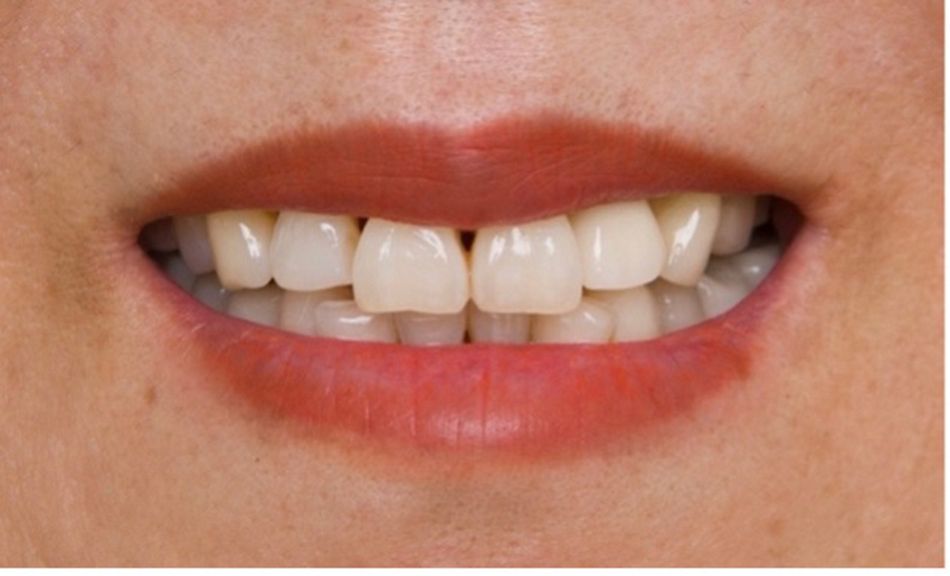
The extraoral examination revealed a high smile line with slightly protruded central incisors and mild gingival recessions with a slight loss of papillae.

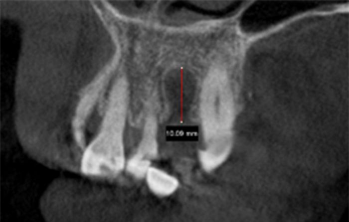
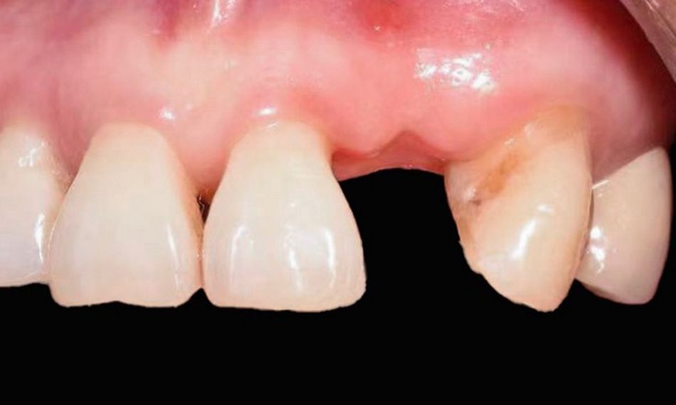
The intraoral examination revealed the absence of tooth #22 and loss of contour of the buccal plate in the area of #22 (Fig. 1). The amount of bone in the edentulous area was determined using CBCT. The CBCT scan confirmed the absence of horizontal bone availability for implant treatment. In addition, there was a vertical defect of 10.09 mm (Fig. 2).

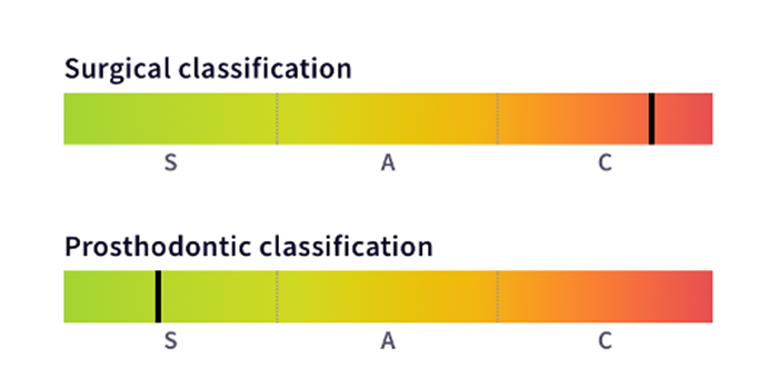
The SAC classification was used as it provides a method for evaluating the potential difficulty, complexity, and risk of an implant-related rehabilitation related to individual implant dentistry cases. It also assists clinicians in the selection of patients, treatment planning and decision-making. This patient was classified as a complex surgical case and straightforward from the prosthodontic standpoint (Fig. 3).

Treatment planning

After discussing the various treatment alternatives, advantages, and disadvantages with the patient, we decided to proceed with a bone augmentation procedure on site #22 and place a Straumann® Bone Level implant after healing. The workflow included the following main steps:

  1. Surgical design of the vertical bone augmentation procedure through CBCT images.
  2. Extraction of tooth #48 and harvesting of the autologous bone block from the zone.
  3. Guided bone regeneration procedure at site #22.
  4. Straumann® Bone Level implant placement 6 months after GBR surgery. 
  5. Healing cap placement 3 months after implant surgery (second-stage surgery). 
  6. Definitive cement-retained crown delivery 2 weeks after second-stage surgery.

Surgical procedure

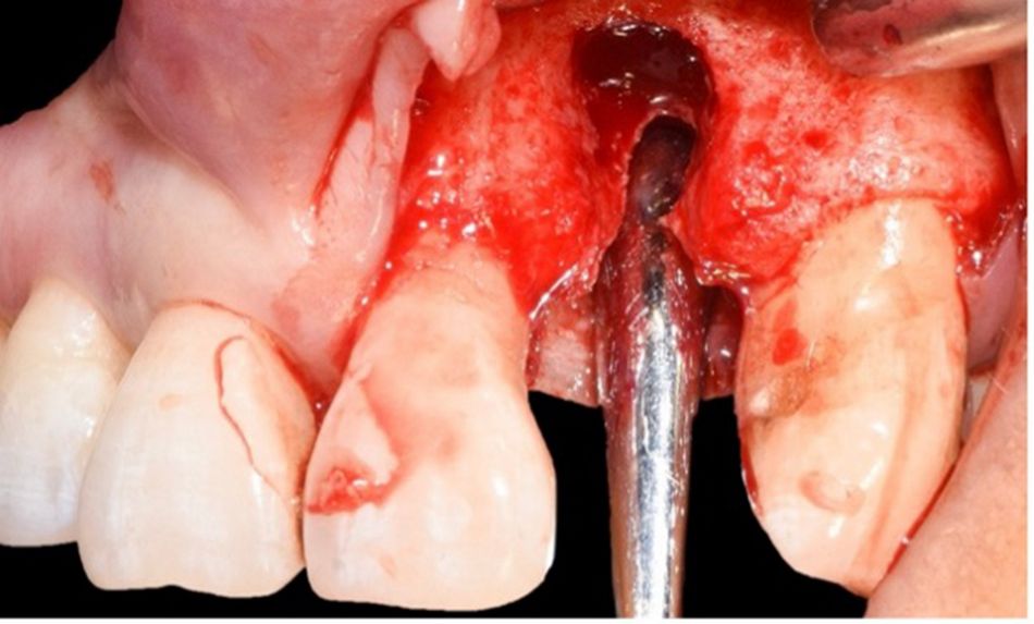
The anesthetic infiltration was done with 2% lidocaine and 1:100,000 epinephrine in the area corresponding to tooth #22. A mucoperiosteal flap was carefully elevated with intrasulcular and crestal incisions. The vertical bone defect was exposed, and the granulation tissue was removed and cleaned until the area was clean and the morphology of the defect could be assessed (Fig. 4).

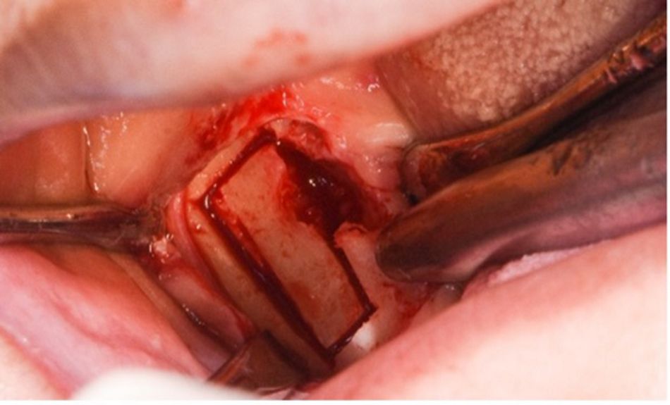
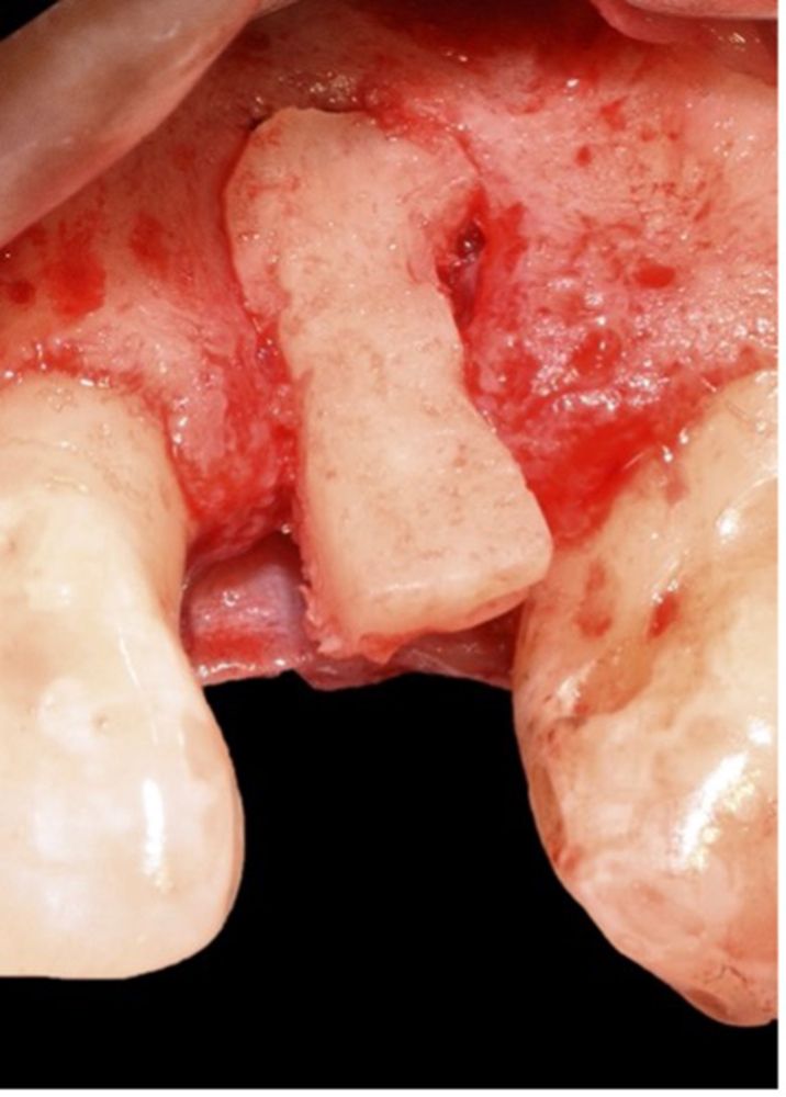
Tooth #48 was extracted in order to obtain an autologous bone block from the zone and regenerate the missing bone in the region of tooth #22 (Fig. 5). Bone harvesting can be used to repair implant sites that have lost bone structure due to a previous extraction.

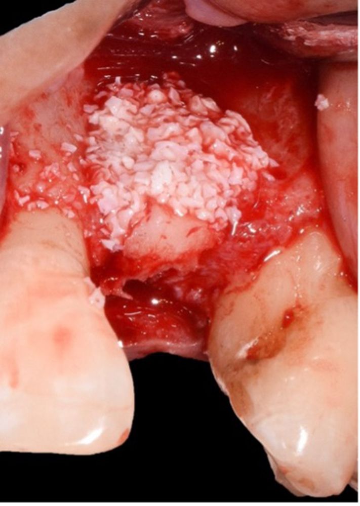
The harvested bone block was shaped to fit the implantation area (Fig. 6). Considering that transplantation should be done within 5 minutes, this step was done in a very efficient manner. The autologous graft was fixed, and xenograft granules were added (Fig. 7). Internal releasing incisions were done to allow a primary closure of the wound.


A Center of Dental Education (CoDE) is part of a group of independent dental centers all over the world that offer excellence in oral healthcare by providing the most advanced treatment procedures based on the best available literature and the latest technology. CoDEs are where science meets practice in a real-world clinical environment.


Sutures were placed and, after two weeks, the patient returned for a follow-up visit. The healing was uneventful, and no complications were reported. The sutures were removed during this visit.

Six months after the GBR surgery, the soft tissue healing was optimal, and the radiographic assessment showed the achievement of 7 mm ridge augmentation. Therefore, the implant placement was planned.

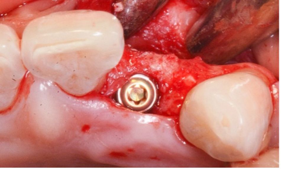
On the day of the surgery, following the anesthesia infiltration and the preparation of the implant bed according to the manufacturer’s instructions, a Straumann® BL ∅3.3 SLA® 10 mm implant was inserted clockwise with the aid of the handpiece at a speed of 15 rpm and torqued to 35 Ncm in the preserved ridge. Primary stability was accomplished (Fig. 8).

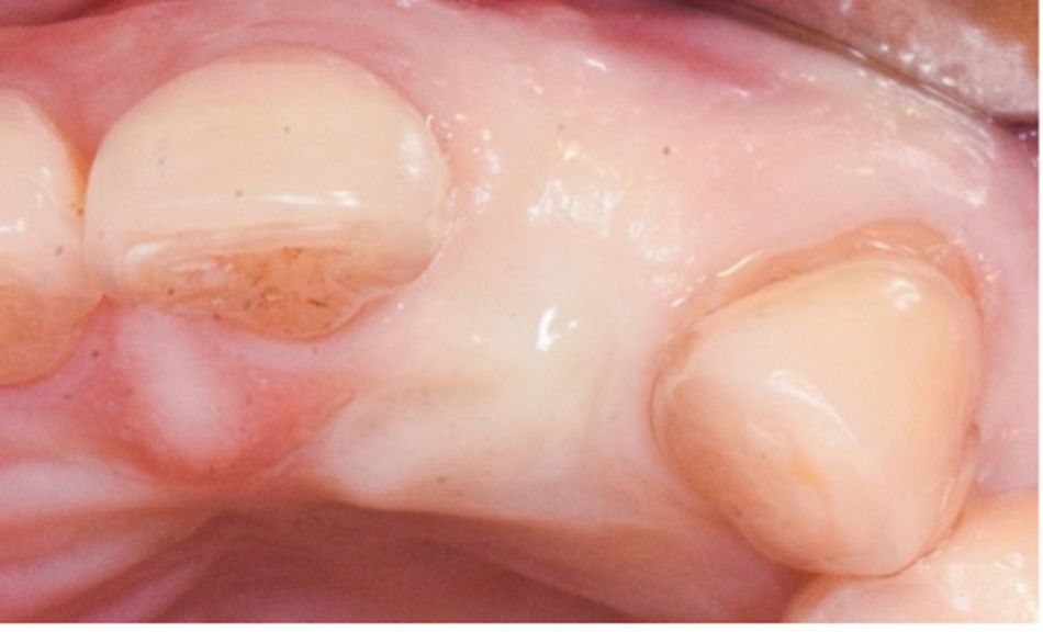
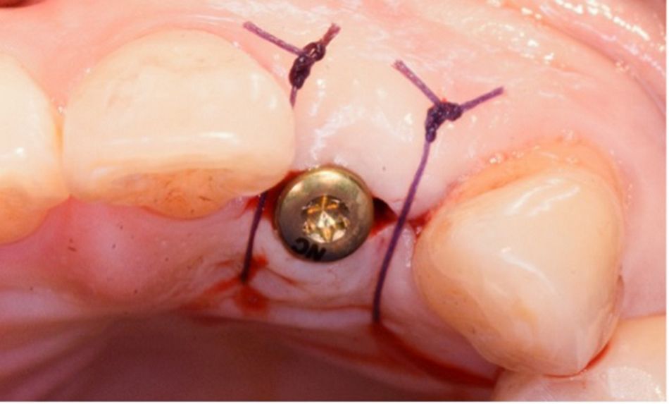
After implant insertion and three months of healing, the implant exhibited good stability with no biological complications. The ridge dimension was 6.5 mm horizontally (Fig. 9). During this appointment, the top of the implant was exposed, and a healing cap was placed (second-stage surgery) (Fig. 10).

Prosthetic procedure

Two weeks after the second-stage surgery, the sutures were removed, and the healing was found to be uneventful. Impressions were taken and sent to the lab to proceed with the preparation of the final restoration.

The cement-retained crown was placed. The occlusion was checked, and oral hygiene instructions were given. The esthetic outcome met the patient’s expectations (Fig. 11).

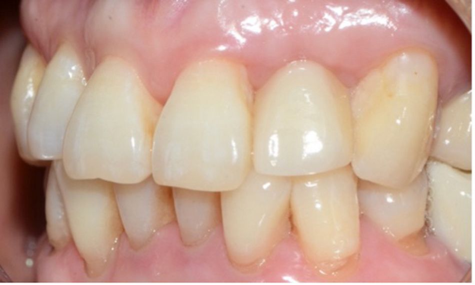
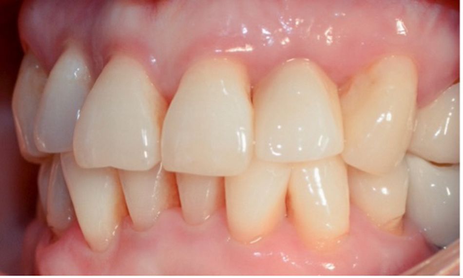
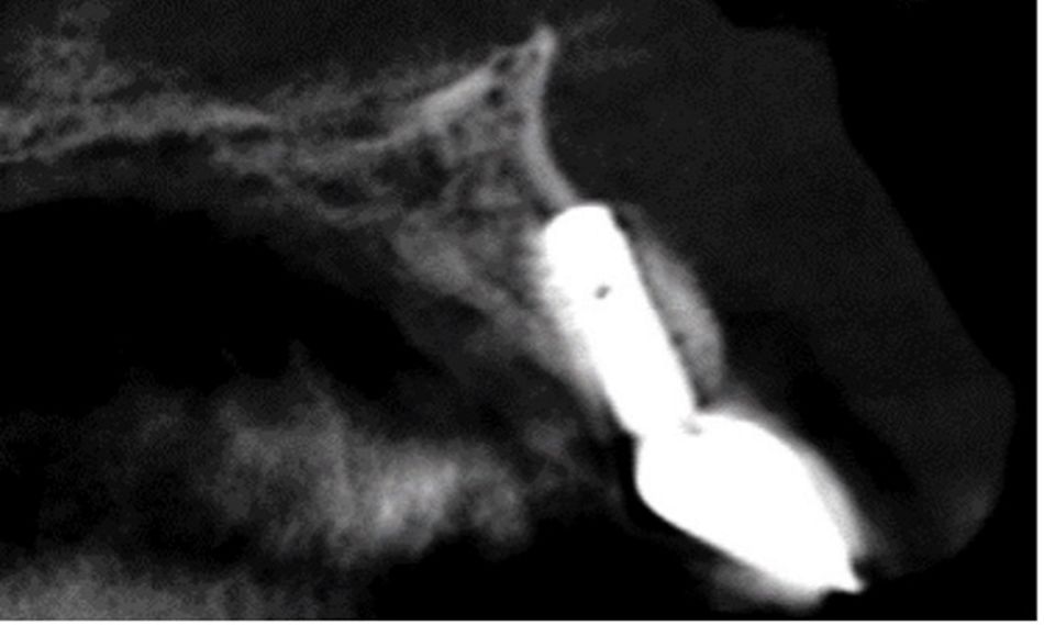
The patient was involved in an annual maintenance program with clinical and radiographic assessments and reinforcement of oral hygiene instructions. After two years, the stability, esthetics, function, and health of the peri-implant tissues were maintained (Figs. 12-14).

Treatment outcomes

The final esthetic and functional outcomes met the patient’s requirements. Furthermore, the peri-implant health was maintained over time. These improved our patient’s quality of life, as she was able to chew and smile with confidence again. During the last follow-up visit, she stated, “I went through a treatment that involved several stages, but it has been worth it; the results exceeded my expectations. The new tooth looks very natural and, in all this time, I haven't had any problems with it.”

Author’s testimonial

“Vertical bone augmentation predictability is substantially lower and the complication rate substantially higher than with horizontal ridge augmentation procedures. When designing the surgical procedure for vertical bone defect reconstruction, it is necessary to understand the bone height of adjacent teeth (adjacent alveolar ridge), because this determines the bone height that can be achieved by bone augmentation.”

References:

  1. Roccuzzo M, Ramieri G, Bunino M, Berrone S. Autogenous bone graft alone or associated with titanium mesh for vertical alveolar ridge augmentation: a controlled clinical trial. Clin Oral Implants Res. 2007 Jun;18(3):286-94.
  2. Buser D, Dula K, Hess D, Hirt HP, Belser UC. Localized ridge augmentation with autografts and barrier membranes. Periodontol 2000. 1999 Feb;19:151-63.
  3. Naenni N, Lim HC, Papageorgiou SN, Hämmerle CHF. Efficacy of lateral bone augmentation prior to implant placement: A systematic review and meta-analysis. J Clin Periodontol. 2019;46 Suppl 21:287-306.
  4. Kumar, G., Parthasarathy, H., Ponnaiyan, D. Localized ridge augmentation with simultaneous implant placement followed by soft tissue augmentation in the maxillary anterior region: A case report. World Academy of Sciences Journal 4.5 (2022): 31