#Immediacy 20. Jan 2017

Replacement of the upper left central incisor with immediate placement and a customized abutment on a Variobase® crown

A clinical case report by Herminio García-Roncero, Spain

Immediate implant placement following tooth extraction might be a viable alternative to delayed placement. However, it requires careful case selection. The implant position and the available peri-implant soft tissue become increasingly important in a patient with a high lip line. This case report describes a surgical technique that preserves anterior esthetics, combining minimally invasive extraction, immediate implant placement and provisionalization, grafting of the buccal space and a connective tissue graft from tuberosity.

Patient history

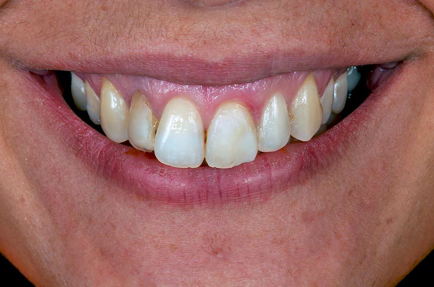
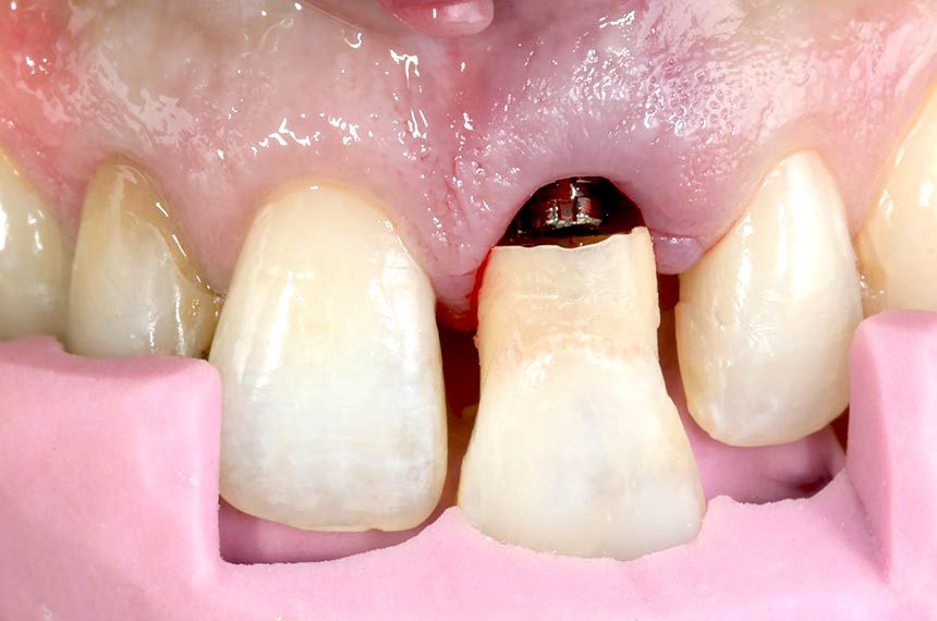
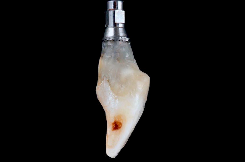
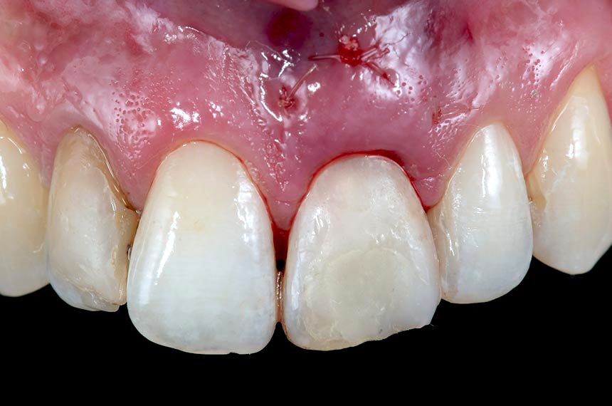
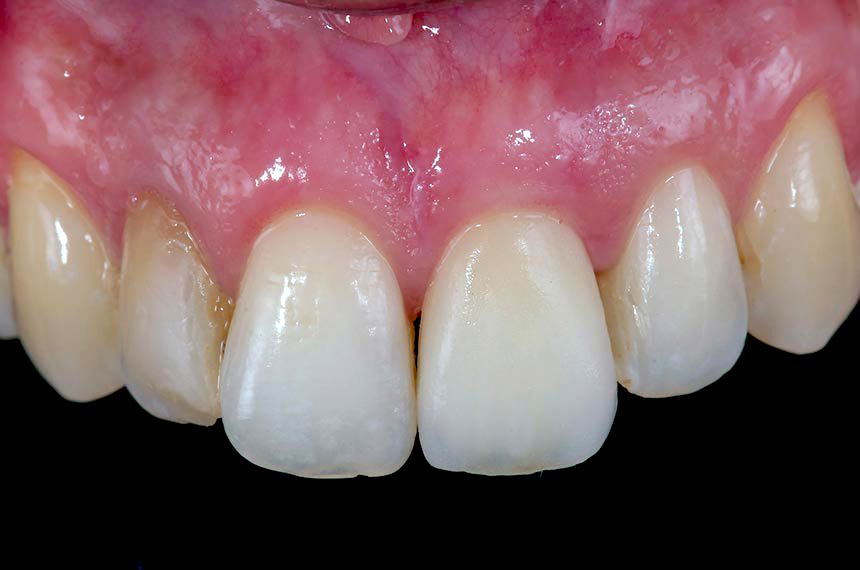
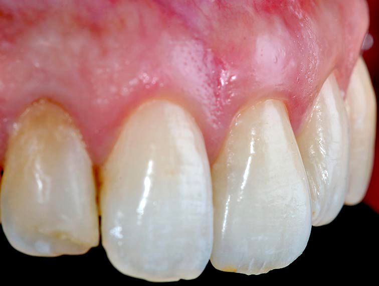
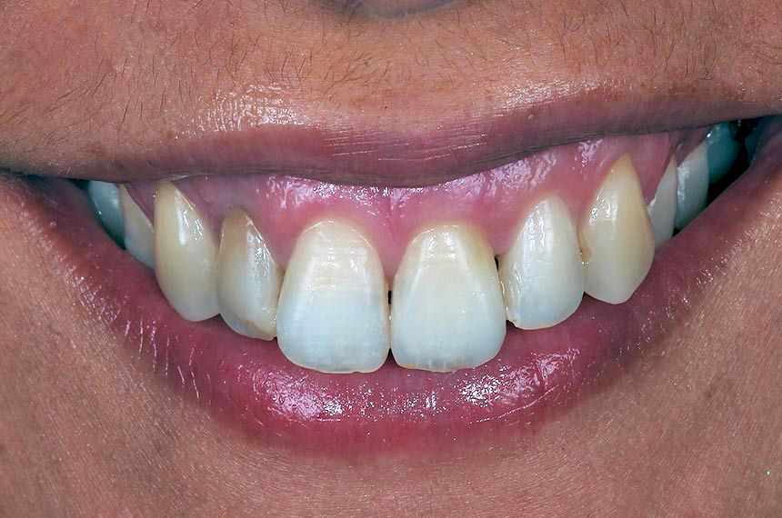
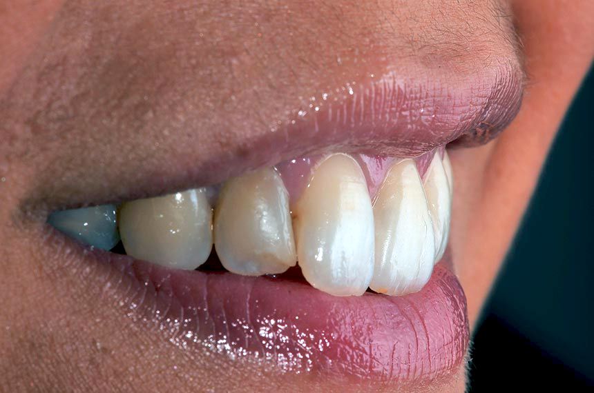
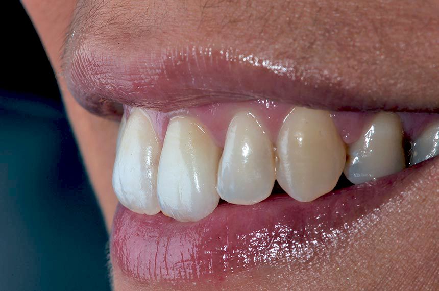
The patient, a 29 year-old Caucasian woman, non-smoker in good general health, was referred to the author’s private dental office with symptoms of vertical root fracture on the maxillary left central incisor (Fig. 1). There was no significant bone loss. The maxillary left central incisor had a history of periapical surgery and endodontic retreatment (Fig. 2). Patient’s oral hygiene was fair. The patient’s history showed no pathological findings and no contraindications with regard to dental implant therapy. The patient has a high lip position that exposes the gingiva so her expectations with regard to the esthetics of the treatment outcome were very high (Fig. 3). It was decided that the maxillary left central incisor should be replaced by an immediate implant.

Surgical procedure

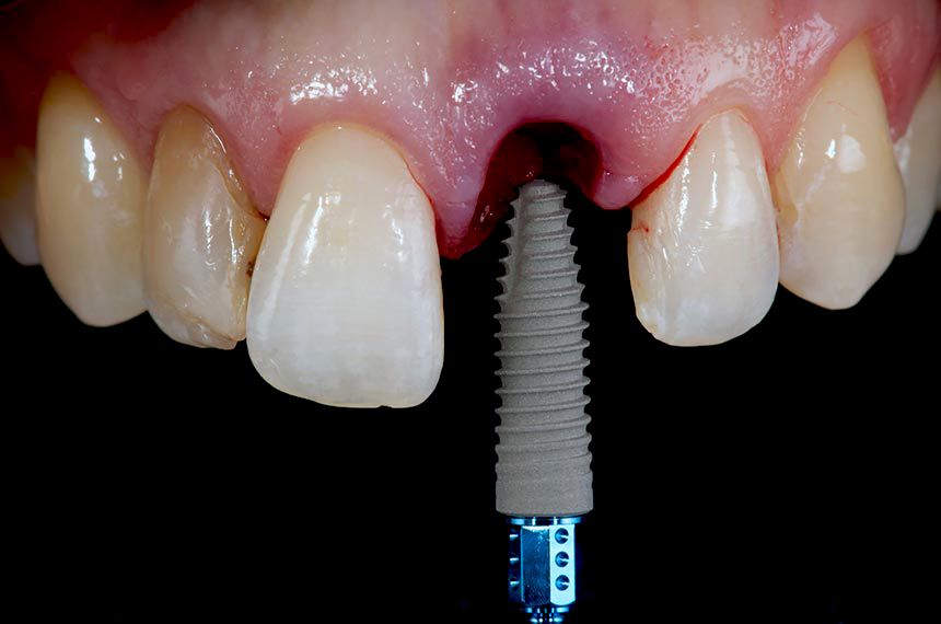
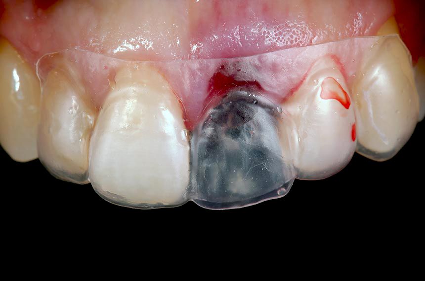
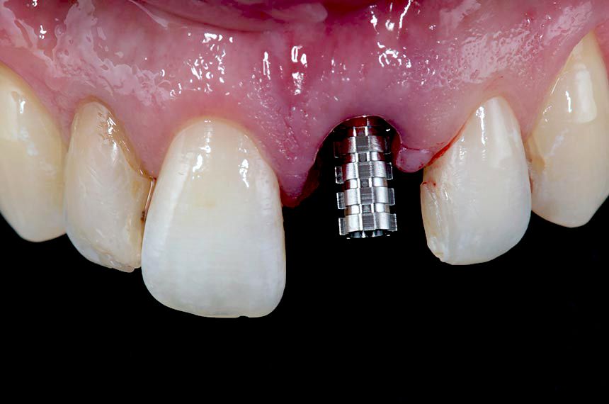
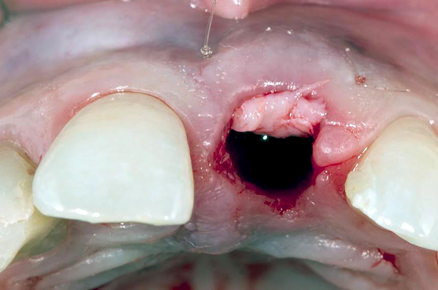
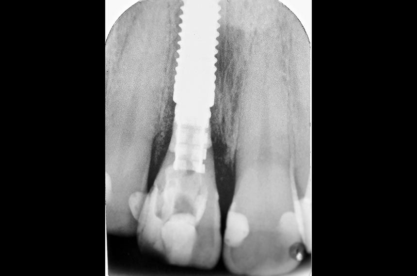
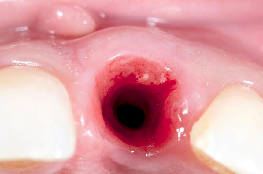
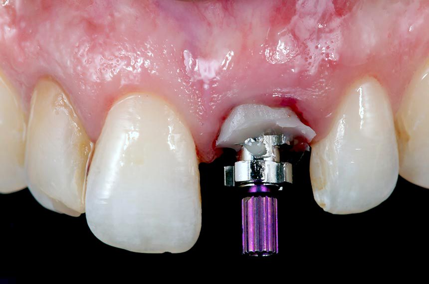
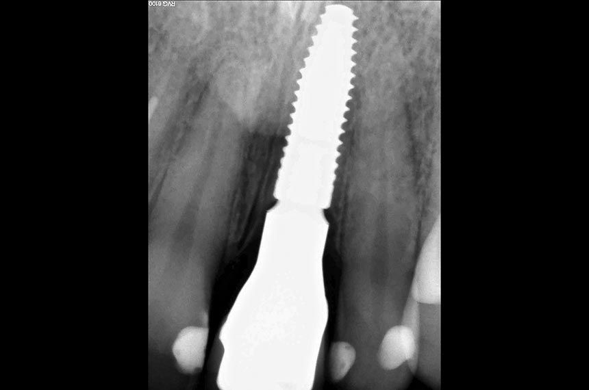
Following administration of local anesthesia, atraumatic tooth extraction was accomplished without flap reflection to preserve the interproximal papillae and the remaining buccal and lingual plates of bone. The extraction socket was carefully examined for dehiscences and fenestrations and debrided of residual periodontal fibers using curettes. Following socket debridement, a Straumann Bone Level Tapered Implant (Regular CrossFit™ Connection Ø 4.1mm, SLA® 14mm) was placed in tooth position 21 (FDI) (Fig. 4). A surgical guide was used to help ensure ideal three-dimensional placement (Fig. 5). The implant showed adequate initial stability when placed with a torque driver at 35 Ncm. A customized provisional titanium cylinder (Temporary Abutment) was then placed and hand-tightened onto the implant and trimmed using a positioned guide to ensure the correct height (Fig. 6). The hollowed crown of the extracted tooth was relined with acrylic resin and adapted to the custom provisional abutment using a positioned guide to ensure exact positioning (Figs. 7, 8). The provisional shell was precisely finished with composite resin on the temporary abutment to recreate the cervical emergence of the extracted tooth (Fig. 9). A synthetic bone graft substitute (Straumann BoneCeramic) was placed between the buccal plate of bone and the implant in order to fill the space and maintain the soft tissue contour. After immediate implant placement and bone augmentation, a supraperiosteal envelope was made labial to the socket. These tunnels, used to accommodate the connective tissue graft, were dissected sharply with a microsurgical blade (Fig 10). Care was taken to leave the periosteum attached to the bone during dissection. A connective tissue graft from tuberosity was inserted into the prepared envelope space and secured with non-resorbable sutures (Fig. 11). The immediate provisional restoration was tightly screwed onto the implant by hand. The provisional restoration was adjusted to clear all contacts in centric occlusion and during eccentric movements. A periapical radiograph was obtained to ascertain the fit of the provisional restoration (Fig. 12).

Clinical situation at the end of the surgery (Fig. 13)

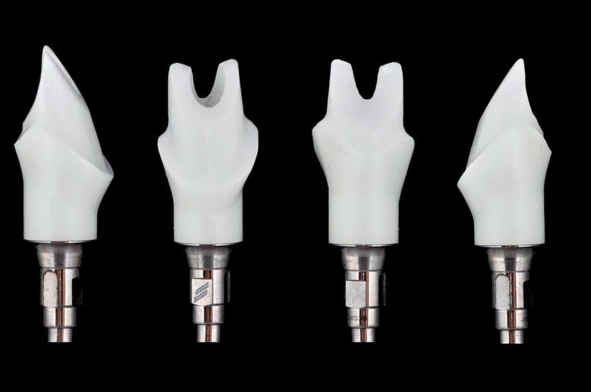
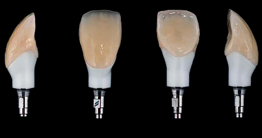
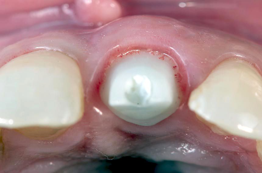
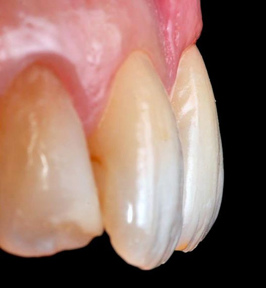
Labial view of the provisional restoration 3 months after the implant surgery (Fig. 14). Note the preservation of the tissue architecture. The provisional restoration was retrieved (Figs. 15, 16). An implant impression was made using a custom impression coping to transfer a record of the healed anatomic tissue to the laboratory (Fig. 17). A definitive customized hybrid abutment on Straumann® Variobase® crown with specific gingival emergence profiles and anatomical shape established by the provisional restoration was fabricated (Figs. 18, 19). The finished abutment was torqued to 35 Ncm, and a provisional PMMA CADCAM restoration was cemented (Figs. 20-22). Three months later a definitive impression was recorded using the lithium disilicate CADCAM core of the definitive restoration as a snap-on abutment coping (Fig. 23). The abutment was never removed during the prosthetic procedures. The definitive restoration was cemented using a resin cement (Figs. 24, 25). The gingival complex surrounding the implant has remained stable. The peri-implant soft tissue was healthy, with no signs of inflammation (Figs. 26-30). The final periapical radiograph revealed no peri-implant bone loss radiographically (Fig. 30).