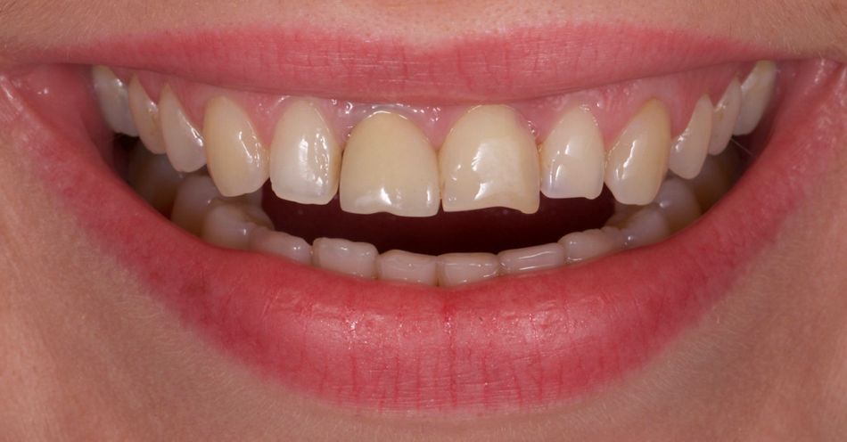
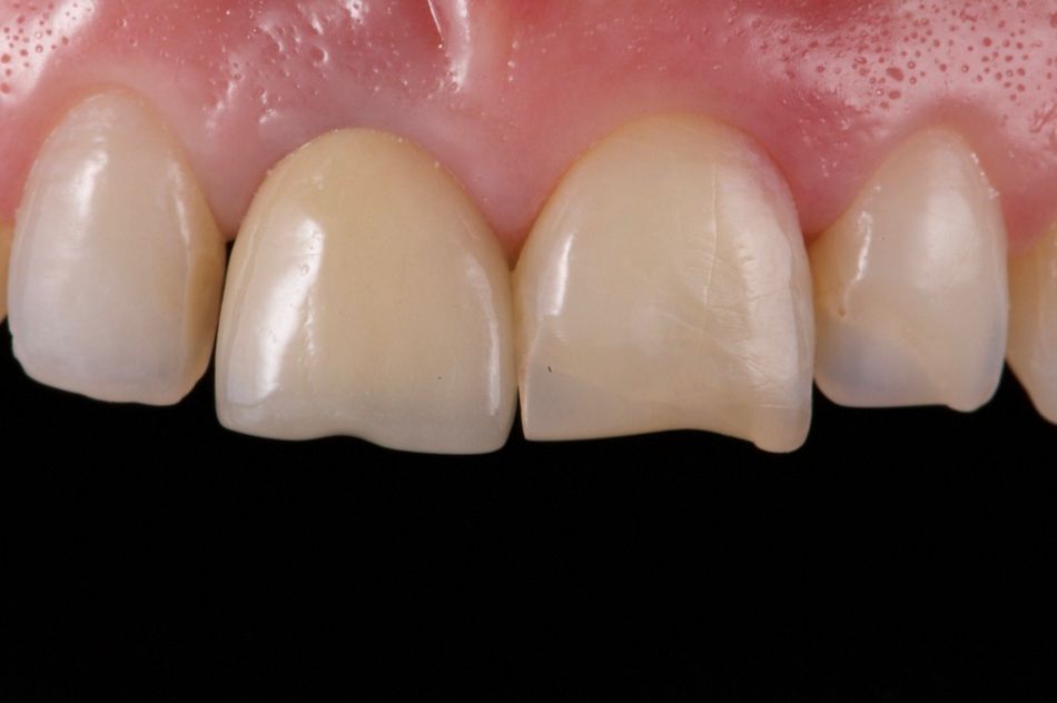
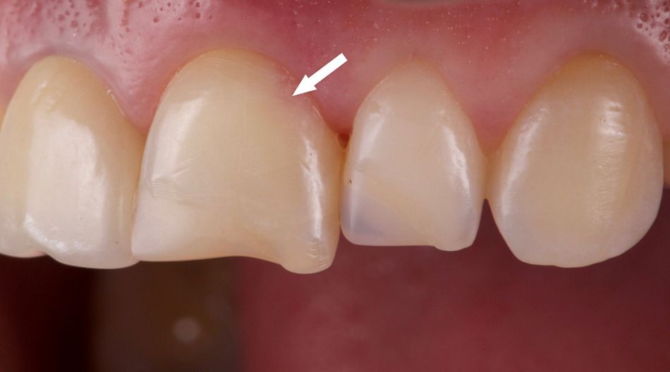
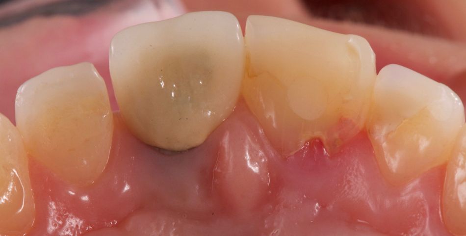
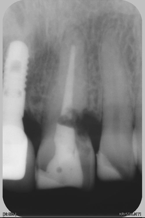
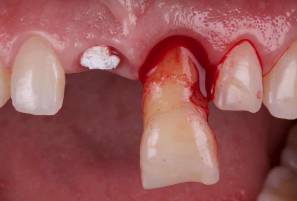
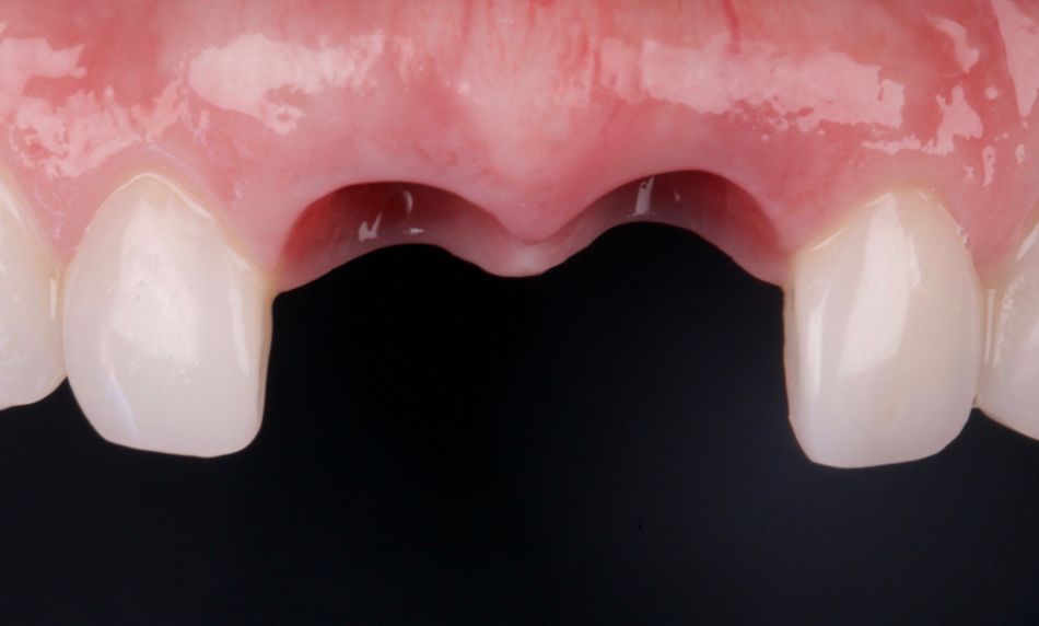
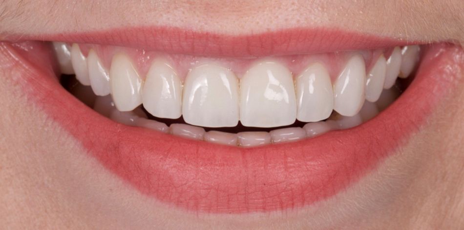
The patient, a 33-year-old non-smoking female with no systemic diseases, presented at our clinic with the request to improve her smile and change the shape, color and inclination of her maxillary anterior teeth (Fig. 1) Clinical examination revealed a PFM crown on the previously placed implant 11, old composite restorations on teeth 12, 21 and 22 (Fig. 2). The pink color of the cervical zone of tooth 21 (Fig. 3) and growth of soft tissue into the tooth crown (Fig. 4) raised suspicion of root resorption. The x-ray examination confirmed the diagnosis of a cervical external root resorption that could not be treated endodontically (Fig. 5).
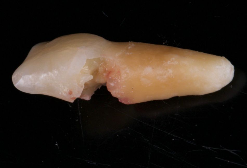
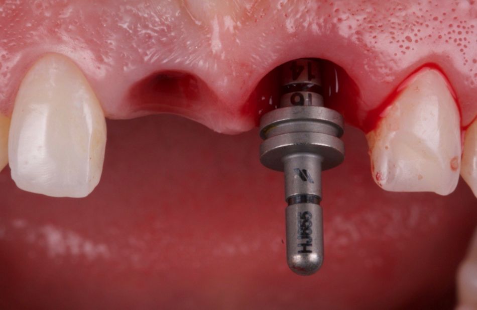
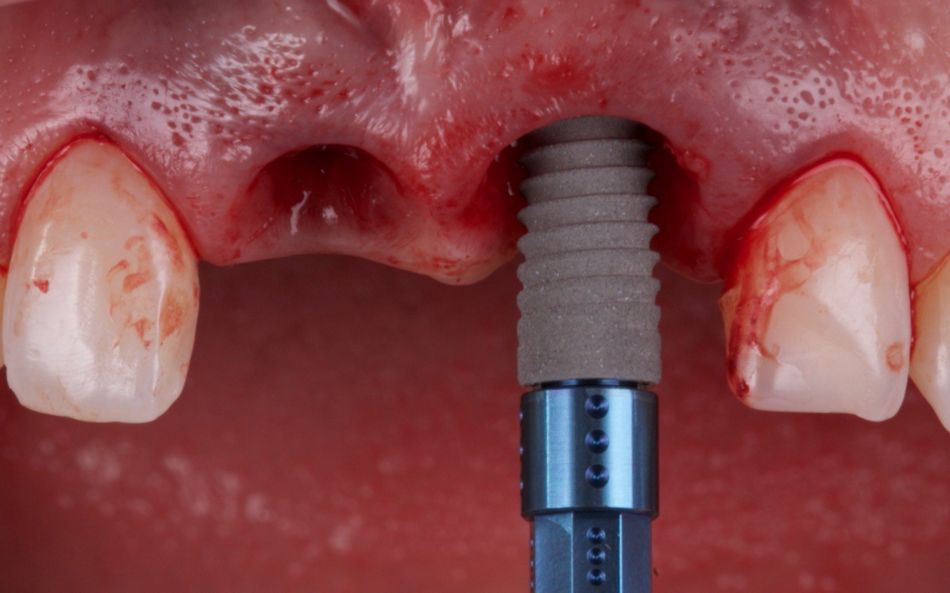
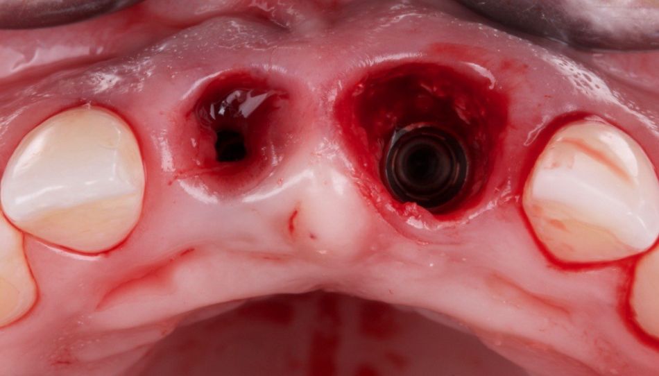
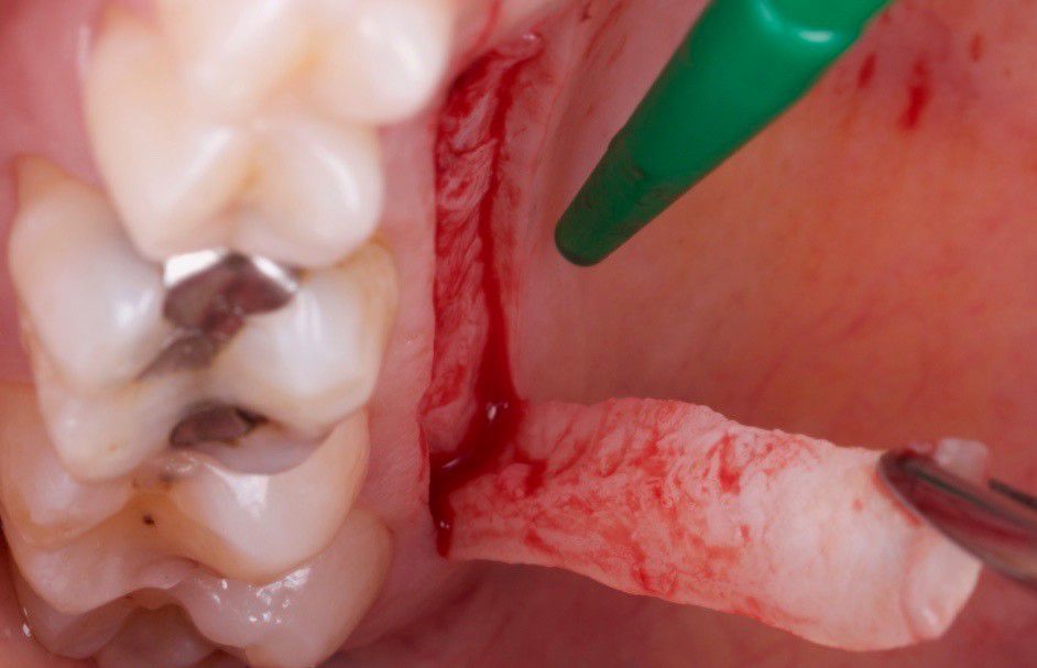
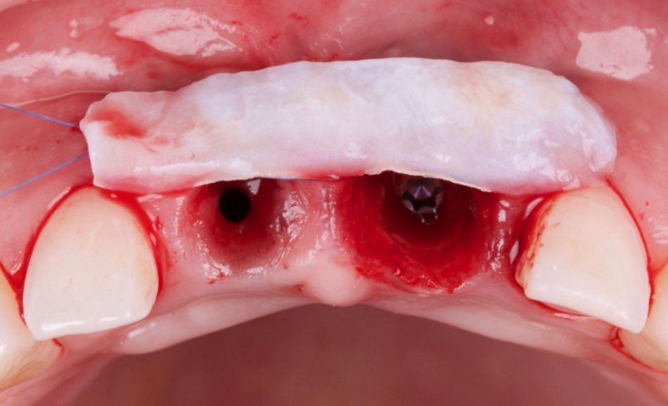
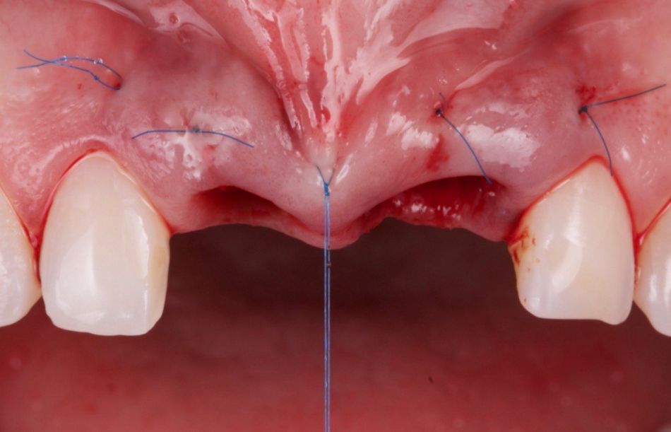
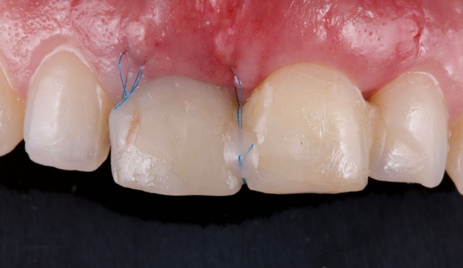
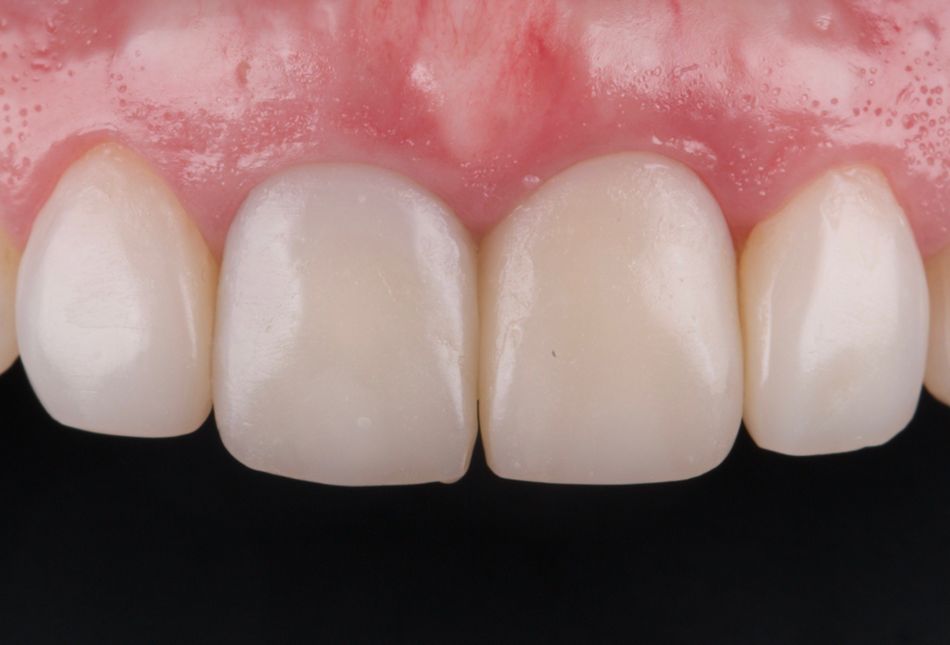
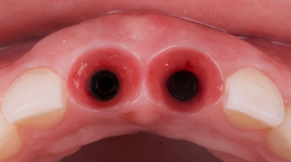
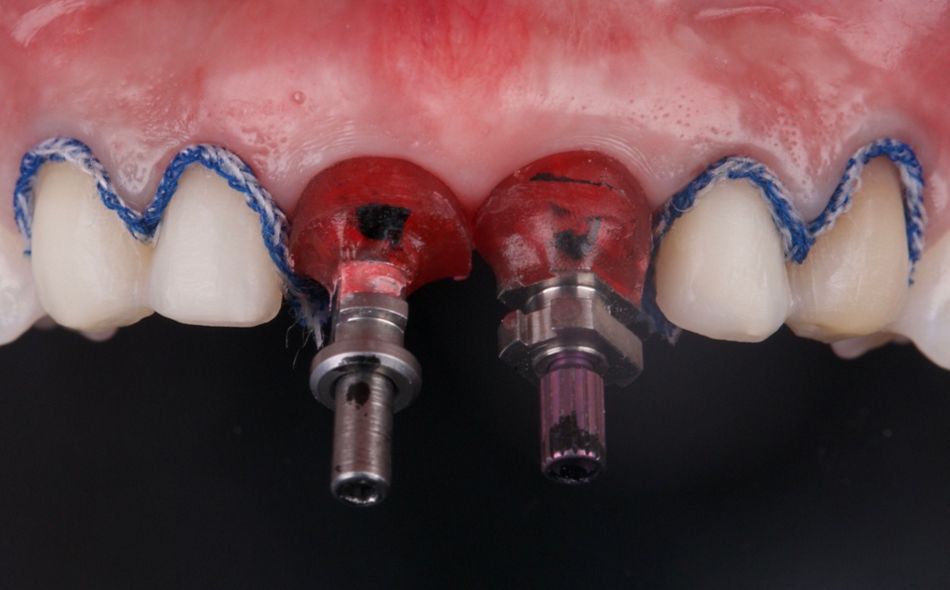
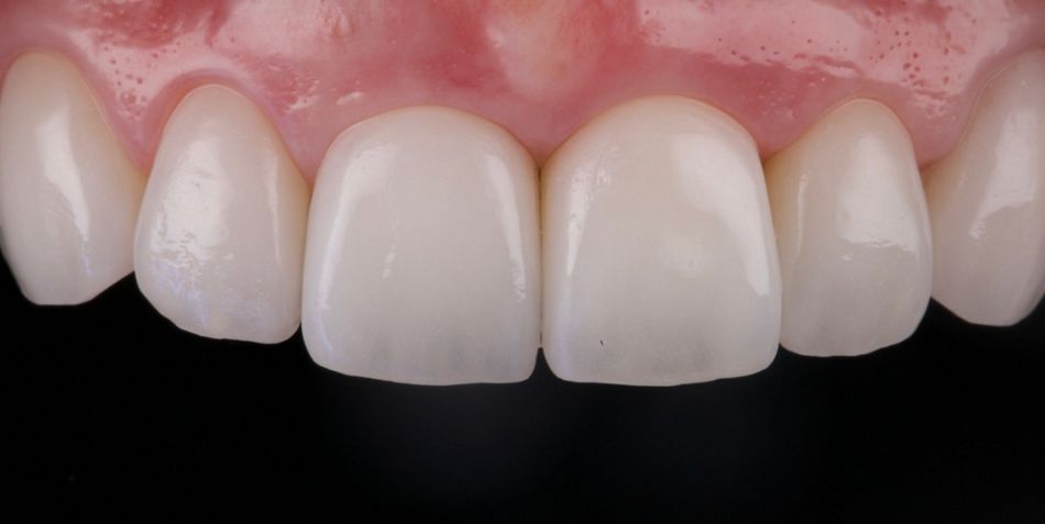
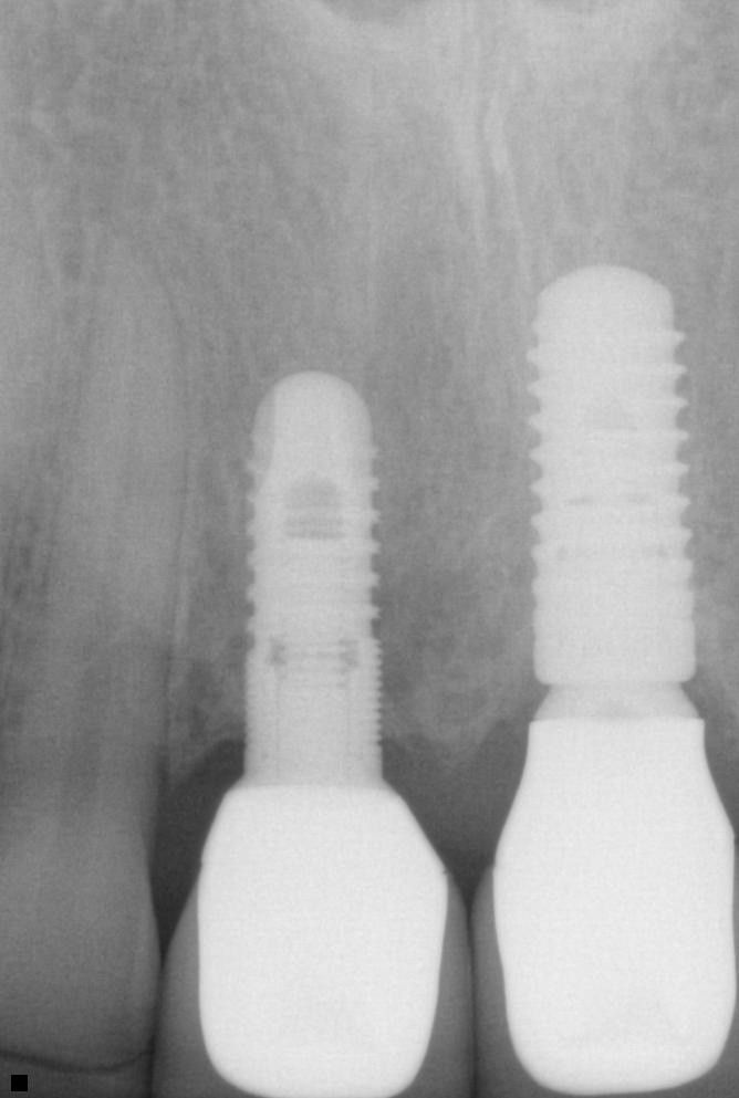
The surgical stage of the treatment consisted of atraumatic extraction of tooth 21 (Fig. 6). We observed a significant root resorption that could not be treated conservatively (Fig. 7). An immediate implant was placed using a Straumann Bone Level RC implant with a diameter of 4.1mm and length of 10mm (Figs. 8-11) In order to increase the soft tissue volume and improve the biotype, a free gingival transplant was placed in the area of the two implants. A free gingival graft was taken from the palate (Fig. 12) and sutured firmly in place (Figs. 13-14). Additional sutures were placed with the support of the temporary crowns (Fig. 15). A second set of temporary implant-supported crowns were made to improve the emerging profile and shape the soft tissues (Figs. 16-18). Impressions were taken using customized open tray impression transfers (Fig. 19). Porcelain veneers were applied to teeth 13, 12, 22 and 23 and the crowns on implants 11 and 21 (Fig. 20). An x-ray (Fig. 21) and the patient’s smile pictures (Fig. 22) were taken two years after placement of the ceramic restorations. The patient was satisfied with this acceptable outcome.
Nowadays, immediate implant placement is a reliable and predictable method that enables us not only to solve and fix the problem with a dental arch defect and solve functional and biological problems, but that also gives us an opportunity to treat the patient in such a way that we do not spoil the smile or quality of life, allowing the patient to continue social activities without major concerns.


