#Immediacy 25. Jan 2021

Immediate rehabilitation of a fully edentulous maxilla with regular implants combined with Straumann® Zygomatic Implants

A clinical case report by Jean Baptiste Verdino, France

A sixty-six years old women presented at the dental office complaining about her sub-complete partial denture (one natural tooth remaining) which showed instability, discomfort and poor esthetic appearance. This denture was in place for 20 years and was considered deficient for more than 15 years.

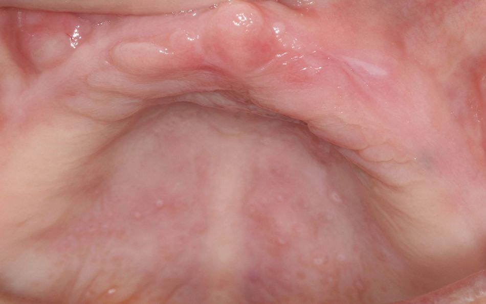
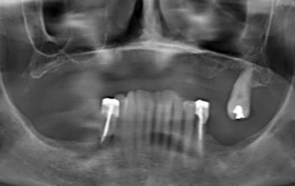
This non-smoking patient was healthy and did not take any medication. Soft tissues were quantitatively and qualitatively in good shape (Fig. 1).  They were no molars at the mandible. The panoramic X-Ray showed a lack of bone at the posterior part of the maxillae (Fig. 2)

Treatment planning

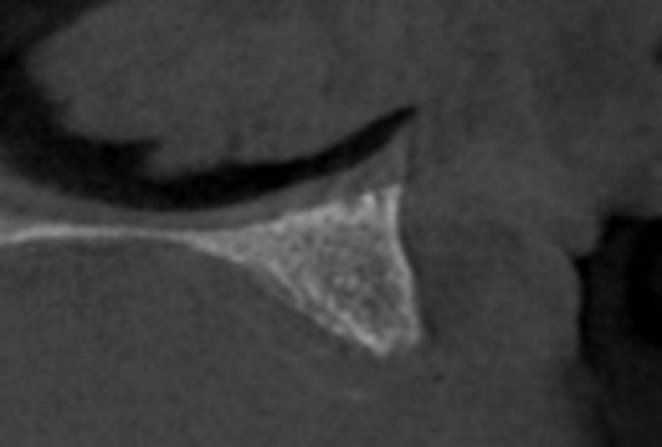
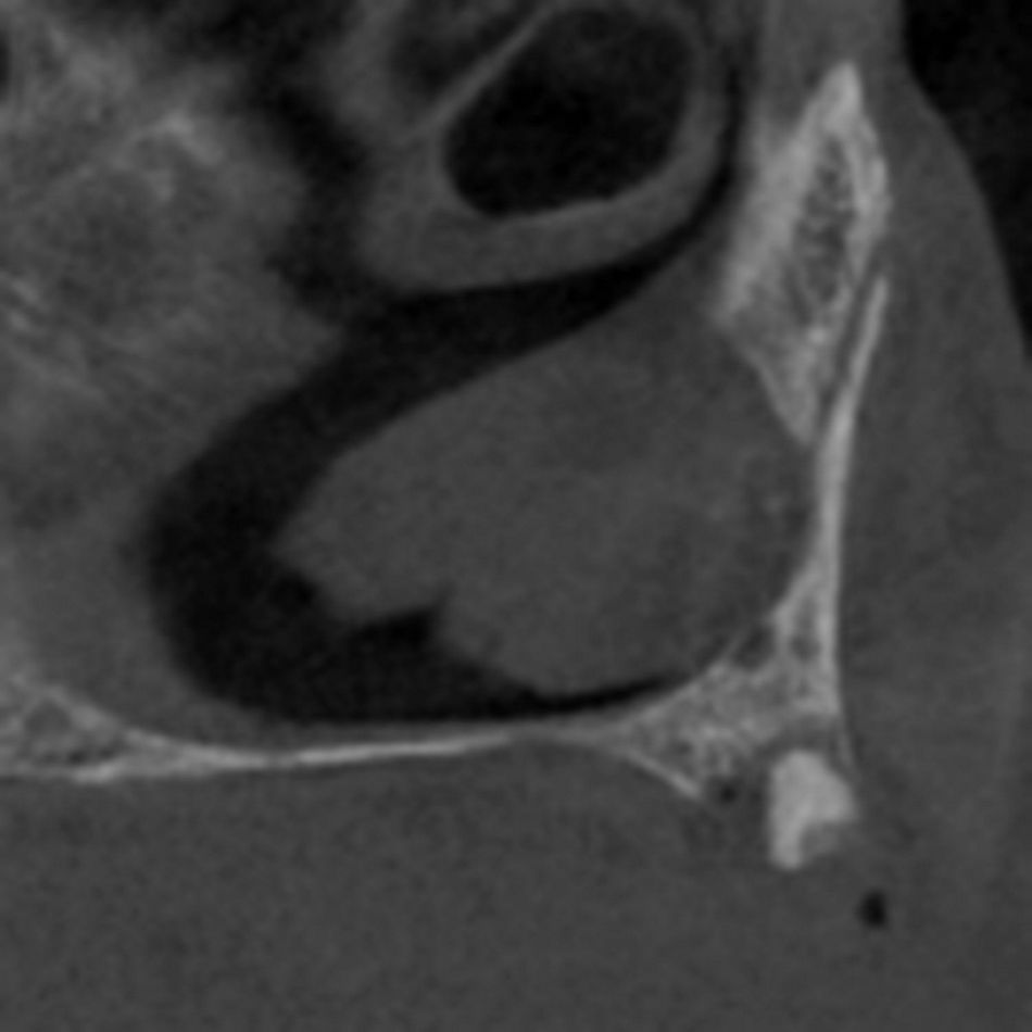
The CBCT showed the presence of remaining bone in the canine region on both sides and confirmed a lack of bone at both posterior parts of the maxilla. A wisdom tooth was foreseen to be removed on the left side, as well as a small piece of root under the mucosa. (Fig. 3,4,5).

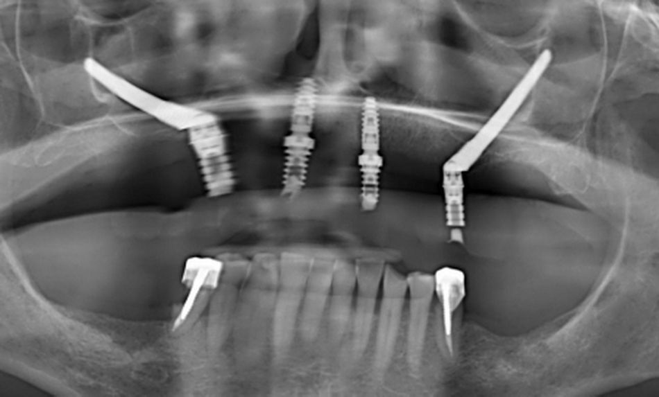
It was decided to place two regular implants in the anterior area, combined with two Straumann® zygomatic implants (one on each side) in order to provide the patient with a fully screwed fixed restoration immediately after the surgery.  The anatomical situation in the posterior area was classified ZAGA 1/2 and thus, two round implants were inserted.

The main advantages of this type of implants are as follows:

  • The small diameter rough surface at the apex of the implants allows to leave enough space available 
  • The machined threaded part underneath the abutment permits to steady an excellent initial stability which is decisive to perform securely an immediate loading.

Surgical procedure
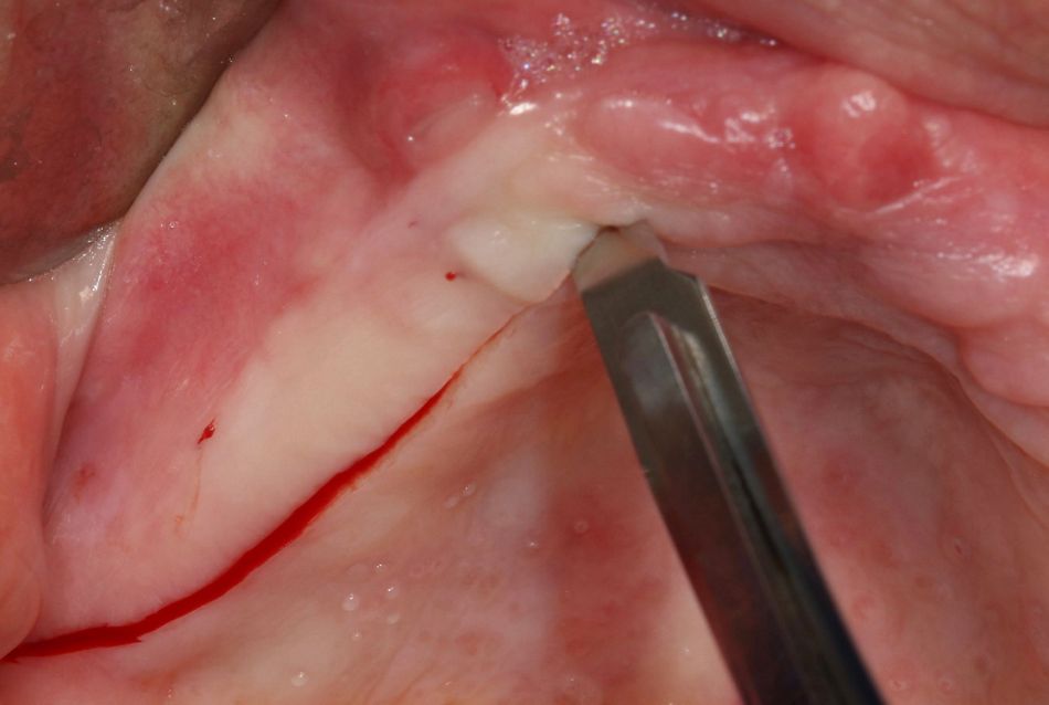
The procedure was conducted under local sedation. The surgery was performed in two phases, firstly right side, then secondly left side, in order to decrease the time of bone exposure to the environment. This is known to facilitate the short-term healing by decreasing the swelling. A large incision (Fig. 6) was drawn, slightly on the palatal side from the top of the crestal bone, in order to dispose later easily the keratinized tissue around the abutment. This incision started from the distal part of the tuberosity, up to the incisive papilla.

An Axiom Anthogyr® PX 3,4/10mm implant was placed in the canine region with an insertion torque of 40 Ncm, allowing for an immediate loading. A regular Multi unit abutment (1,5mm/4,8mm) (Anthogyr, Sallanches, France) was screwed at 25 Ncm on the top and covered with a healing cap.

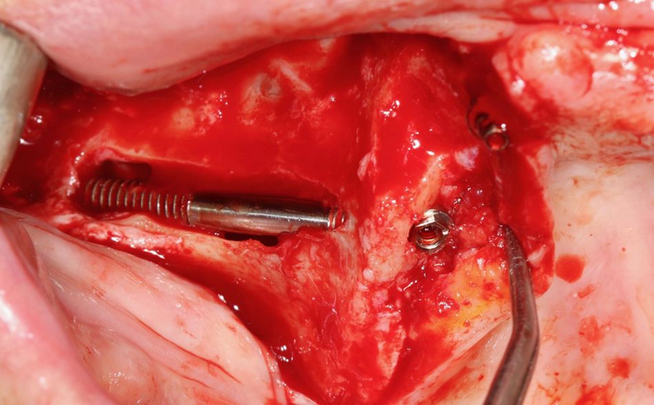
For the placement of the zygomatic implant and according to the ZAGA 1/2 classification, a long window was performed at the anterior wall of the sinus, in order to place the head of the zygomatic implant as close as possible to the remaining alveolar crest. This improves the comfort for the patient and eases future cleaning procedures. The classical drilling procedure, using a round bur and a single drill was performed and the implant (Straumann® Zygomatic Implant Zaga round, length= 40mm, Institut Straumann, Basel, Switzerland) inserted and tightened at 50Ncm (Fig. 7).

It is important to notice that the head of the implant must be fully surrounded by bone providing two major advantages:

  • High level of initial stability.
  • The bone around the implant will allow for a better gingival health and prevent future inflammation.

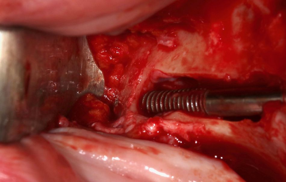
Another important parameter for surgical success is to be able to visualize the emergence of the apex of the Zygomatic implant at the buccal face of the zygomatic bone. Thus, the surgeon can be sure that the implant is well in place (Fig. 8)

A Straumann® multi-unit abutment 1,5 mm (Institut Straumann, Basel, Switzerland) was placed, tightened at 35 Ncm and covered with a healing cap. Resorbable sutures were used to close the flap and particular attention was paid to  the soft tissue management around the abutment resulting in  a thick amount of keratinized gingivae positioned around the abutment.(Fig. 9)

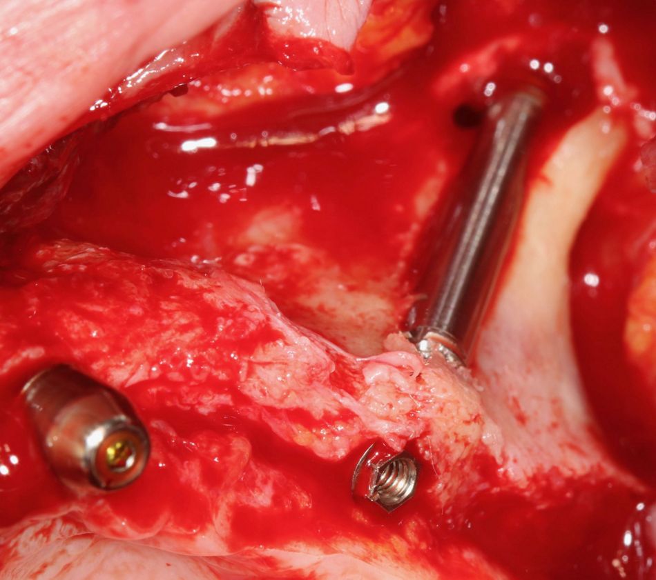
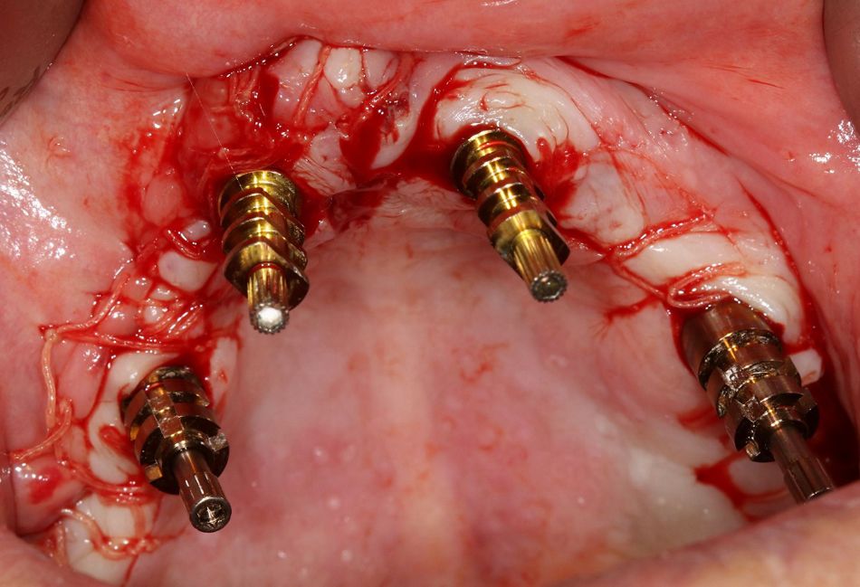
The second phase of the surgery followed the same pattern. A regular implant placed in the front (Anthogyr® Axiom PX 3,4/10, Multi unit abutment 1,5mm/4,8mm), an a second Straumann® Zygomatic implant Zaga round 40 mm (Institut Straumann, Basel, Switzerland) with 1,5mm Multi unit abutment placed posteriorly. As for the right side, the anatomical situation (ZAGA1/2) allowed for the placement of a cervical emergence close to the top of the bone crest with surrounding bone around the implant (Fig. 10).

The initially elevated flap was closed with resorbable sutures.

Prosthetic procedure

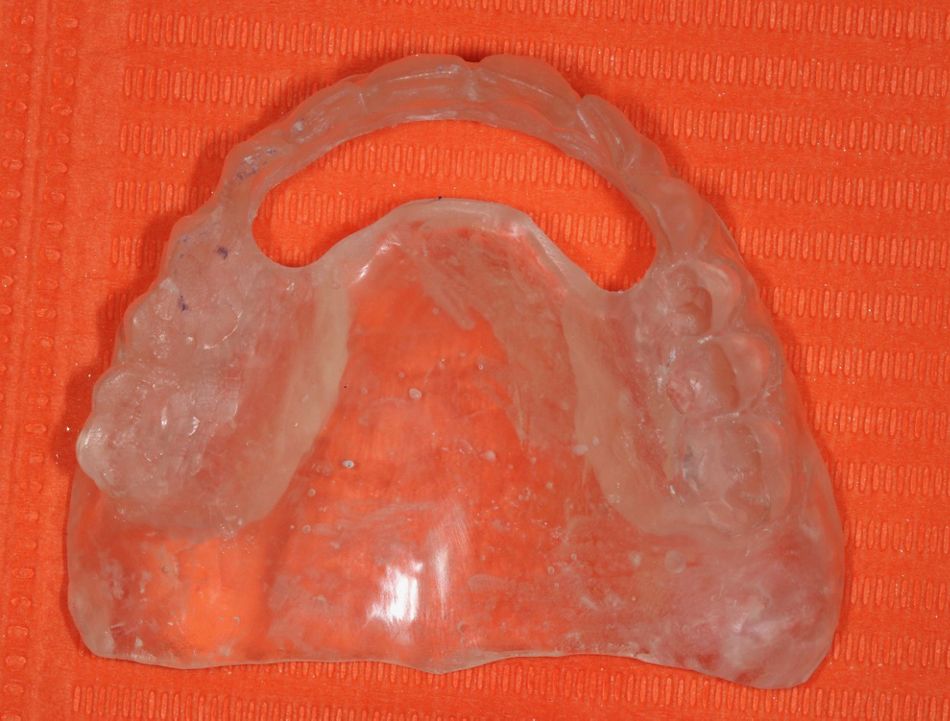
At the second appointment, an aesthetic try-in was validated by the patient in order to obtain his agreement on the shape and shade of the teeth. This enables to manufacture a translucent resin bite rim, by simple duplication of the try in. (Fig. 11)

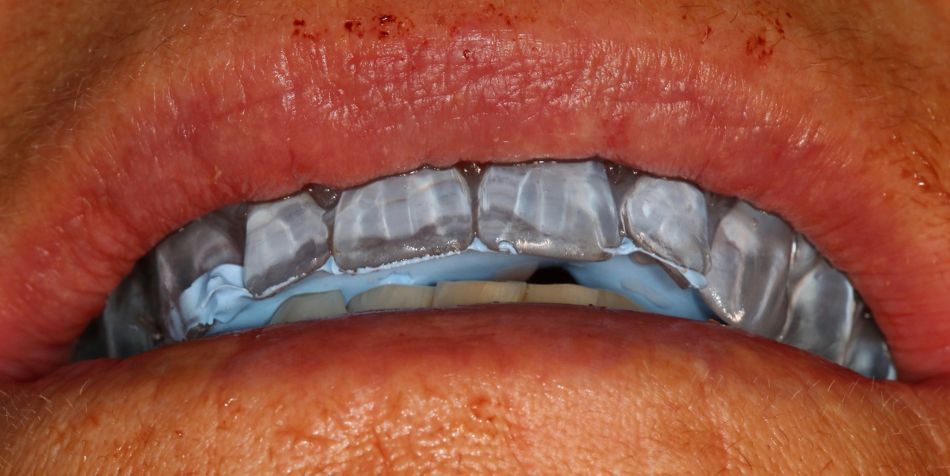
This approach authorizes the occlusal registration, by filling the resin bite rim  with bite registration paste (Blue Bite Coltene®, Henry Schein Inc., USA) to stabilize it, before placing the bite material in between the two jaws. (Fig. 12)

The exact position of the implants is registered with a plaster impression, using screwed transfers and an open tray (Fig. 13)

Laboratory procedure.

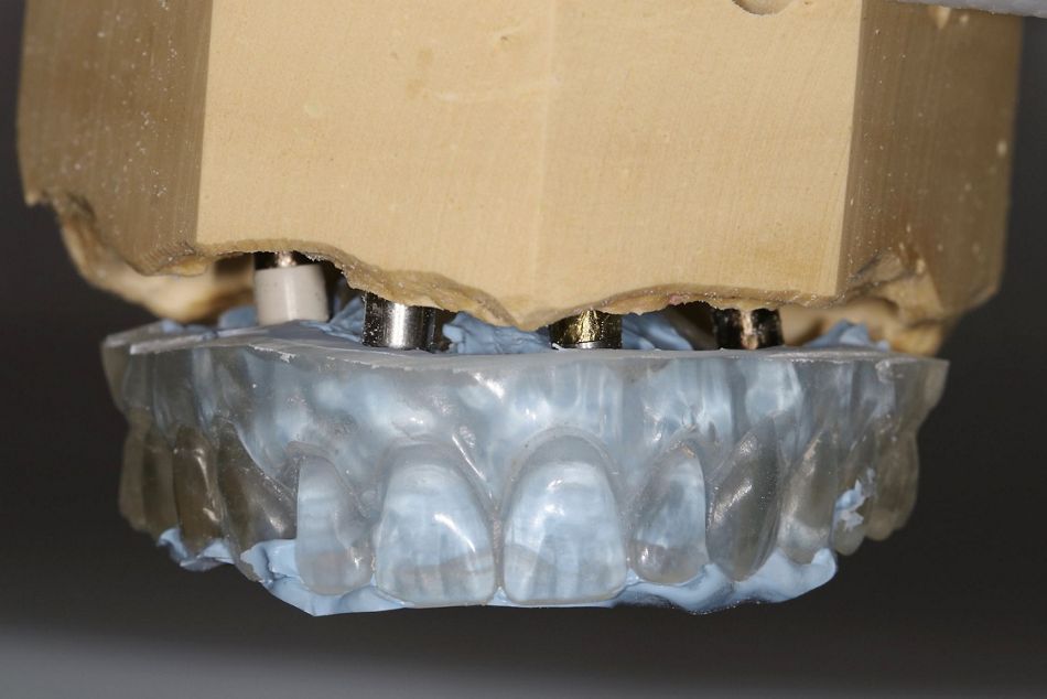
The master model is casted in stone plaster, mounted on a semi-adaptable simulator by use of the template (Fig. 14)

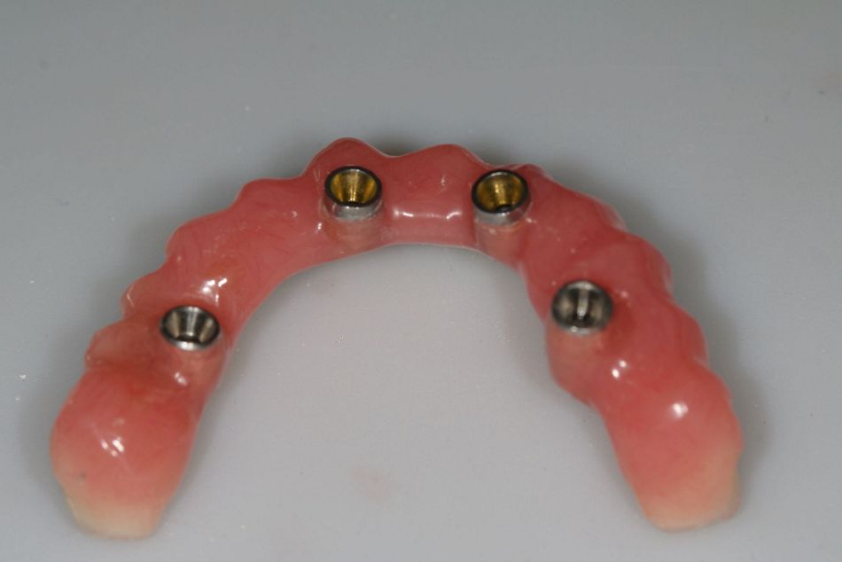
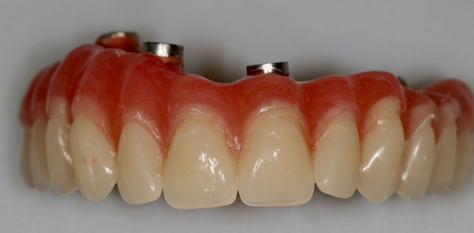
The pre-existing acrylic bridge is connected to the temporary titanium cylinders, previously covered with silane to improve the adhesion of the PMMA to the titanium. The bridge must be smooth and convex in all directions. Avoiding any concave zone hinders plaque retention and ease the hygienic maintenance both parameters which contribute largely to the long-term prosthetic success.

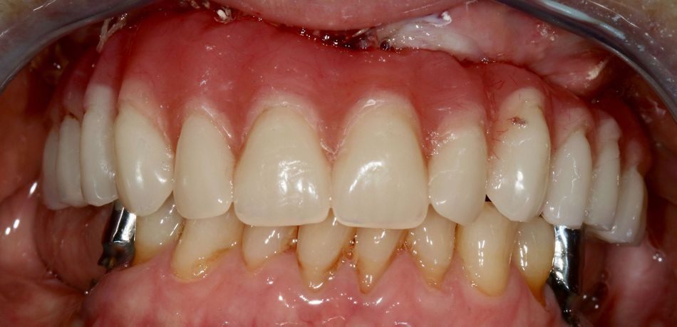
Approximately 3 hours after the end of the surgery, the bridge is screwed in place and the occlusion controlled, as well as the ability for the patient to reach every part of it with an inter-proximal brush. (Fig. 17)

Follow-up instructions regarding chewing (avoid hard food, “eat only what you can cut with a fork”) are outlined and four appointments are scheduled to double-check the healing process.

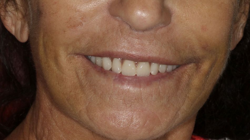
The patient was invited to enjoy her “new smile” and she expressed a high level of initial satisfaction (Fig. 18)

A control panoramic X-Ray was performed and confirmed the accurate placement of the implants and supra-structures.

The author acknowledges Mr Jean Michel Moal, Dental Prosthetic Technician at Verdino Clinic for his outstanding contribution.