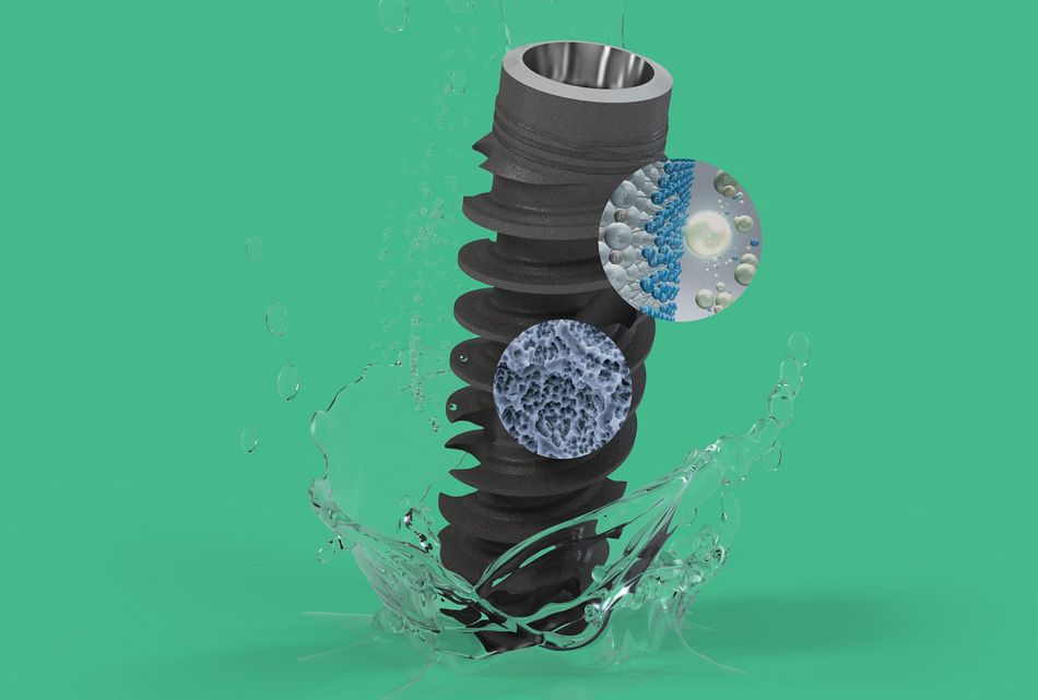
- The success of dental implants depends on strong and long-lasting bone-to-implant integration. Modern research highlights the pivotal role that immune modulation plays in the early stages of osseointegration, where cells like neutrophils and macrophages orchestrate the body’s healing environment. A delicate balance between pro-inflammatory (M1) and regenerative (M2) macrophages remains essential for optimal outcomes, with the timely polarization process being key to reducing chronic inflammation and promoting predictable bone regeneration.
- Surface characteristics of implants at macro, micro, and nano levels can significantly influence such responses, fostering the development and maintenance of a pro-healing environment in the peri-implant tissues. Notably, advanced surface technologies like SLActive® exemplify how immune system modulating properties, including enhanced hydrophilicity and nanoscale topography of the dental implant surface, can facilitate early healing as well as stable and predictable osseointegration, particularly in complex or high-risk cases.
- As our knowledge of immunobiology continuously advances, the development of contemporary implant surfaces, such as SLActive®, has established a new benchmark for predictability and long-term success in implantology.

Introduction
When it comes to the success of dental implants, few factors are as critical as effective bone-to-implant integration – or, in other words, osseointegration. Initial investigations into osseointegration occurred accidentally in the 1950s when Prof. PI Branemark observed that a titanium chamber he had implanted into rabbit bone for another study had become integrally bonded to the bone. This discovery provided foundational insights into the biological compatibility of titanium with bone tissue, pivotal for the advancement of implantology (1, 2). While the process of osseointegration is often seen as a straightforward bone response, mounting research suggests a more nuanced view: it is an immune-driven process. Therefore, it's important to understand that osseointegration isn't a passive bonding process. Instead, the success or failure of the development of a tight and long-lasting bond between bone and implant relies heavily on a biological response process known as immune modulation (3, 6).
Role of immune cells in osseointegration of dental implants
To understand how the immune responses work, we should take a closer look at what happens after implant placement. When a dental implant is placed in the bone, the body initiates an inflammation-driven response where key immune cells play a central role in determining the outcome of the integration process (4, 6). These cells don’t, however, act alone. Instead, they engage in critical crosstalk with each other and the bone. This “signaling” determines whether the local environment supports healing and successful bone formation or enters into a “fight or flight” response dominated by prolonged inflammation, the latter of which can greatly impair the osseointegration process and eventually lead to implant loss (5, 6). The first immune cells to respond upon implantation are neutrophils. Their function involves triggering an initial inflammatory response that protects the area and helps prepare the site for healing (6, 7, 24, 28). Macrophages - a secondary responder, have a more complex role; they manage the healing process. These adaptable immune cells possess the ability to shift between two functional states – or phenotypes, based on the surrounding microenvironment. Essentially, M1 macrophages promote inflammation, helping to protect the environment by controlling the possibility of early infection. In contrast, M2 macrophages trigger anti-inflammatory activity, tissue repair, and bone regeneration (8). The ability to switch between these two phenotypes – a process known as macrophage polarization is essential in dictating the direction of the tissue healing process, hence implant osseointegration (9, 10). While an M1 response is necessary for initial defence, a prolonged M1 state can hinder bone healing, thus increasing the risk of implant failure. Conversely, when macrophages commence a timely shift toward an M2 phenotype, they create an environment that reduces inflammation and promotes the recruitment of bone-forming cells (osteoblasts) to begin bone formation around the implant (11). Therefore, effective osseointegration depends on achieving proper immune modulation, where M1 macrophages provide essential early protection, and M2 macrophages are critical for advancing toward bone healing and osseointegration. Any delay or inadequate transition between these stages can compromise the stability and longevity of the implant. As our understanding of immunobiology and how it affects the osseointegration process continuously deepens, it opens the door to exciting opportunities like optimizing immune response through biomaterial design and implant surface modification. This allows for achieving more predictable long-term treatment outcomes, especially in difficult or complex cases (12, 19, 29, 30, 34, 48, 49, 50).
Implant surface characteristics and modulating immune responses
Today, implant surfaces play a key role in the biological performance of dental implants. An implant’s surface topography influences not only integration but also early host immune responses (13, 14, 21). Macro-topography, defined by implant contours and thread geometry, visible to the human eye, is designed to enhance primary stability and facilitate mechanical interlocking with the supporting bone. However, it’s at the micro and nano levels where implant surface characteristics interact with and influence cellular and molecular mechanisms. Micro-roughness, typically in the range of 1-10 µm, promotes cell adhesion but also increases the available surface area, thus creating a favorable microenvironment for bone matrix deposition (14, 41, 42). The nano topography, generally characterized by dimensions smaller than 100 nm, is not found on all implant surfaces. This feature was first identified and thoroughly documented on the Straumann® SLActive® surface. The SLActive® implant surface builds upon the proven success of sand-blasted, large-grit, acid-etched, (SLA) technology (44, 45, 46) by incorporating surface chemistry designed to initiate and enhance early biological responses (20, 47). Unlike conventional hydrophobic surfaces, SLActive® exhibits a high surface energy and a strong affinity for protein and blood adhesion. This hydrophilic property, coupled with micro- and nano-structured surface topography, plays a pivotal role in modulating the early immune response, a critical factor of implant integration (14, 15, 16, 21). The nano-roughness found on the SLActive® surface comprises TiO2-based ultra-fine structures, which are formed during a hydrothermal treatment step following the SLA process surface treatment (17, 21, 52). It has been confirmed that the nano-scale surface topography profoundly impacts protein accumulation, which in turn, influences the recruitment and behavior of immune cells (14, 15, 16). Moreover, nano-rough surfaces have often been associated with enhanced hydrophilicity, improving protein adsorption and early cell interactions. This is important because hydrophilic surfaces reduce the invasion of neutrophils while enhancing favorable cytokine (signaling) profiles to create a ‘pro-healing’ microenvironment. These advances in surface engineering and hydrophilicity not only enhance osseointegration but also contribute to reducing the risk of chronic inflammation and early implant failure (13, 14, 17, 21). Understanding and controlling these immune responses is central to the next generation of dental implants. SLActive®’s wettability and nanoscale surface characteristics influence the conformity and bioactivity of proteins, leading to improved cell attachment, recognition and signaling. Importantly, this optimized interface reduces the activation of pro-inflammatory neutrophils and promotes the early recruitment of M2 Macrophages (23, 28). These features have also been shown to influence macrophage phenotype polarization helping them to make the timely shift from a pro-inflammatory M1 phenotype to a regenerative M2 phenotype (4, 9, 39). This shift in immune phenotype is essential for fostering a pro-healing microenvironment that supports early bone matrix formation and timely osseointegration.
What about the other surfaces?
While other implants might provide an acceptable mechanical interlocking capability, their surfaces very often do not present the same degree of immune modulatory properties as SLActive® does. Compared to other commercially available surfaces, both hydrophobic and hydrophilic, Straumann® SLActive® consistently demonstrates an enhanced ability to positively influence immune responses. This was evidenced in a series of studies that directly compared various commercially available implant surfaces in terms of macrophage polarization and pro- and anti-inflammatory cytokine release in vitro. They verified that SLActive® delivers an optimal immunomodulatory effect, which can subsequently lead to faster wound healing and more efficient osseointegration. (4, 9, 39). Animal studies, which addressed the speed and effectiveness of osseointegration, followed by those performed in humans, further confirmed these results (18, 20, 22, 27, 31, 32, 36, 47). The superior early healing response and the reduction of inflammatory burden make SLActive® a highly efficient and predictable tool for both initial integration and long-term success (35, 51).
Practical applications of SLActive® in clinical settings
The surface technology of SLActive® represents a significant advantage in implant dentistry, particularly in clinical scenarios that require high predictability and efficiency. In this context, numerous preclinical and clinical studies have confirmed that implants with the SLActive® surface exhibit faster and more robust osseointegration. (18, 20, 22, 25, 26, 27, 36). Moreover, they are associated with higher survival rates in high-risk patient populations such as those with diabetes, undergoing irradiation, and smokers (12, 19, 29, 30, 34, 48, 49, 50). Due to its ability to facilitate superior bone-to-implant contact (BIC) during the initial healing phase, SLActive® is considered a preferred choice for immediate implant placement and loading protocols, where accelerating the development of secondary stability through enhanced healing is critical to success (33, 35). This is especially relevant in esthetically demanding zones, where obtaining early secondary stability is often necessary. Essentially, SLActive® implants contribute to a smoother learning curve for new implant dentists by minimizing biological complications during the early stages of healing (20, 47). For anyone beginning their journey in implant dentistry, the use of SLActive® implants offers distinct advantages by simplifying treatment protocols, increasing predictability and enhancing clinical outcomes. The optimal immune response modulation, hence reduction of healing time associated with SLActive®, improves patient satisfaction while reducing chair time, allowing clinicians to achieve better results with greater confidence. Moreover, high success rates with immediate and early loading protocols mitigate the need for extensive surgery, thereby reducing procedural complexity. Adopting SLActive® technology enables dentists to broaden their scope of implant treatments, offering solutions for more complex or challenging cases, while maintaining a high standard of patient care and long-term success.
Take-aways
- Effective immune response is a critical determinant of successful osseointegration.
- The physicochemical properties of the implant surface play a critical role in influencing immune cell behavior.
- The SLActive® surface illustrates how advanced implant surface technology can modulate the early immune response to facilitate healing and accelerate osseointegration.
