Immediate full-arch implant rehabilitation using Straumann BLX™ implants within the Straumann Pro Arch™ concept in an atrophic maxilla and mandible
Introduction
Dental implant rehabilitation in patients with severe maxillary and mandibular atrophy represents one of the most demanding challenges in oral rehabilitation. Reduced bone volume and systemic compromise, such as osteoporosis and controlled hypertension, limit the biological potential for osseointegration and increase surgical risk. Careful patient selection and biologically sensitive protocols are therefore essential to achieve predictable outcomes in medically compromised individuals (1).
In contemporary full-arch rehabilitation, digital smile design has become an important adjunct to conventional diagnostic and surgical planning, especially when esthetic integration and facial support are critical treatment objectives. In this case, Smilecloud was incorporated as a digital step to project the intended final restorations and define the esthetic endpoint before surgery. This facially driven preview supported communication between clinician, technician, and patient, while reinforcing a prosthetically driven approach to implant positioning and restorative design.
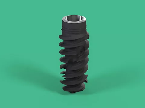
The Straumann BLX™ implant system, integrated within the Straumann Pro Arch™ concept, offers macro-geometry and surface characteristics designed to meet these demands. Its Roxolid® titanium-zirconium alloy provides enhanced tensile and fatigue strength compared with conventional pure titanium, minimizing invasiveness in compromised bone (2). In addition, the SLActive® surface accelerates early bone healing and increases bone-to-implant contact, enabling predictable immediate loading even in challenging conditions (3,4). These properties align with consensus recommendations that support immediate loading when sufficient primary stability is achieved (5).
This treatment approach also emphasizes prosthetically driven planning and the use of Straumann® screw-retained abutments (SRAs) to achieve predictable implant positioning, efficient prosthetic design, and restoration of esthetics and function with reduced treatment time. The addition of Smilecloud at the planning stage strengthened this workflow by allowing the restorative outcome to be visualized in advance and translated into surgical and prosthetic decisions, as well as patient confidence and engagement. This approach is particularly valuable in elderly patients with compromised health, as immediate full-arch fixed rehabilitation minimizes surgical stress, shortens recovery, and provides the comfort and confidence of a stable, functional smile from the very first day (2,6).
This clinical case report describes the use of the Straumann Pro Arch™ concept with Straumann BLX™ implants in the full-arch rehabilitation of an edentulous patient with atrophic ridges and systemic compromise. It illustrates how adherence to consensus guidelines, combined with advanced implant design, digital restorative projection, and biologically sensitive protocols, can overcome anatomical limitations, restore esthetics and function, and significantly improve quality of life in elderly patients.
Initial situation
Patient Background
A 71‑year‑old female patient, with a medical history of osteoporosis and well‑controlled hypertension, and no smoking habits, presented to the clinic seeking a definitive solution after decades of struggling with removable prostheses. For nearly 20 years, she had relied on partial removable dentures in both the maxilla and mandible, which had progressively lost stability and comfort. The patient reported increasing dissatisfaction with her oral condition (Figs. 2a-c), expressing a strong desire for an implant‑supported fixed prosthesis that could restore function, esthetics, and her overall quality of life.
Fig. 2. Digital teeth arrangement using Smilecloud
The patient had high expectations regarding improvements in function, esthetics, and overall oral health. The remaining dentition was severely compromised due to extensive restorations, as well as significant bone and soft tissue loss resulting from periodontal disease.
Clinical Findings
Clinical examination revealed extensive compromise of the remaining dentition. In the maxilla, the remaining teeth showed inadequate spacing, with loss of proximal contacts that had led to food impaction and recurrent inflammation. Extensive restorations were present, some failing with secondary caries and marginal leakage contributing to the overall deterioration of the dental structures.
The long‑term use of partial removable dentures had left its mark: mucosal irritation and evident mucositis were observed in the palatal region, caused by continuous pressure of the prosthesis against the delicate mucosa. This chronic trauma not only resulted in erythematous and inflamed tissue, but also contributed to the patient’s discomfort and reduced tolerance to removable prosthetic wear (Fig. 3a).
In the mandible, the posterior regions revealed pronounced ridge atrophy, with flattened and resorbed crests that posed a significant challenge for implant placement. The loss of vertical and horizontal bone dimensions reflected the cumulative effects of long‑standing periodontal disease and the mechanical trauma of removable prostheses. This resorption had not only compromised the available bone volume, but also altered the anatomical contours, limiting prosthetic support and stability.
The anterior mandible presented an even more critical finding: the edentulous ridge, long deprived of teeth, had evolved into a sharp “knife‑edge” contour. This morphology, resulting from extensive resorption over many years, has significantly complicated the surgical planning. The narrow, pointed ridge offered limited surface area for implant anchorage and increased the risk of cortical perforation, demanding careful bone vertical bone reduction and delicate positioning of implants to achieve stability without compromising safety (Fig. 3b).
The remaining dentition was severely compromised, with extensive restorative work showing recurrent failures, and marginal leakage. Several teeth exhibited mobility and periodontal pockets, underscoring the progression of chronic periodontal breakdown. Gingival recession and loss of keratinized tissue further accentuated the compromised esthetic and functional prognosis.
This constellation of factors reinforced the urgency of a definitive solution. The patient’s motivation for a fixed implant‑supported rehabilitation was not only esthetic and functional, but also deeply emotional, with her desire to overcome years of discomfort and instability, and to regain confidence through a stable, functional smile.
Figs. 3 Intraoral initial situation.
Radiographic evaluation
Radiographic evaluation, including panoramic imaging and Cone Beam Computed Tomography (CBCT) scans, confirmed the markedly reduced bone volume in both arches (Figs. 4a-d). The images revealed extensive resorption patterns consistent with the clinical findings, highlighting the severity of the anatomical compromise. In the maxilla, the loss of proximal contacts and posterior ridge atrophy were evident, while in the mandible the posterior resorption and the anterior knife‑edge ridge underscored the complexity of the case.
These radiographic findings reinforced the lack of a predictable prognosis for conventional prosthetic rehabilitation. Without a complex restructuring supported by a full‑arch implant solution, the patient’s functional and esthetic expectations could not be met. Thus, the evidence pointed decisively toward a comprehensive implant‑supported approach, where the Straumann‑term oral health.
Figs. 4a-c Radiographic initial situation.
Despite these anatomical limitations, the patient’s motivation and the availability of advanced implant solutions made her a suitable candidate for full‑arch rehabilitation using the Straumann Pro Arch™ concept with Straumann BLX™ implants.


Treatment planning
Treatment planning began with the definition of the intended restorative outcome rather than the surgical intervention itself. To establish a facially driven endpoint, Smilecloud was incorporated as a digital smile design and restorative visualization step, allowing the team to project the final restorations according to the patient’s facial features and esthetic expectations, compared to her initial situation (Fig. 5). This digital preview helped define key parameters such as tooth display, smile arc, midline orientation, incisal position, and overall dentogingival balance. By visualizing the desired outcome in advance, the surgical and restorative strategy could then be developed in a fully prosthetically driven manner.
Fig. 5 Smilecloud facially driven comparison between the initial situation and the projected restorative outcome in the upper arch.
Following this planning phase, the clinical condition of the remaining teeth and supporting tissues confirmed the need for full-arch extraction and implant-based rehabilitation.
The next step involved the extraction of all remaining teeth in both arches. These teeth were severely compromised by extensive restorations, recurrent caries, and periodontal breakdown, and their removal was essential to eliminate infection sources and prepare the oral environment for implant placement.
Following extractions, the implant placement strategy was carefully defined. Five Straumann BLX™ implants were planned for the maxilla and four for the mandible. Their distribution was designed to optimize support for a full‑arch fixed prosthesis, despite the presence of posterior ridge atrophy and the knife‑edge anterior mandibular ridge. In the mandible, a controlled bone reduction in the anterior region was planned to transform the sharp ridge into a broader platform, thereby creating sufficient sagittal bone thickness for safe implant anchorage. In the posterior regions, tilted implants were selected to take advantage of the available bone, minimize cantilevers, and achieve a more favorable distribution of occlusal forces. These decisions are consistent with established protocols for full‑arch type 2 rehabilitation, where bone reduction and posterior angulation are critical to achieving stability and long‑term success (5).
The choice of a type 2 full‑arch prosthesis was determined by several anatomical and esthetic factors. According to Misch’s bone classification, the patient presented advanced resorption patterns that limited the prognosis for conventional prosthetic rehabilitation without complex restructuring (7). The knife‑edge anterior mandibular ridge required reduction to achieve adequate width, while posterior bone loss necessitated angulated implant placement. In addition, the patient’s smile line and esthetic demands required a prosthetic design that could compensate for soft tissue loss, harmonize gingival contours, and restore facial support. Together, these considerations made a Ttype 2 prosthesis the most predictable solution, balancing surgical feasibility with esthetic and functional outcomes.
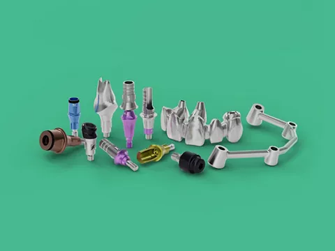
The choice of abutments was guided by prosthetically driven planning. Straight SRAs were designated for axially positioned implants, while 30‑degree SRAs were selected for tilted implants. This ensured predictable implant positioning and facilitated the design of screw‑retained restorations, reducing prosthetic complexity and enhancing long‑term stability.
An immediate loading protocol was incorporated into the plan, following the Straumann‑arch prosthesis was scheduled to be delivered on the day of surgery, restoring esthetics and function without delay. This approach was particularly valuable for the patient, minimizing surgical stress and providing immediate comfort and confidence.
After a healing period of approximately four months, definitive prostheses were planned: a monolithic zirconia prosthesis with buccal veneering in the maxilla, and a titanium‑acrylic prosthesis in the mandible. Both would be screw‑retained to the SRAs, ensuring durability, stability, and long‑term success.
Ultimately, the treatment plan was tailored to the patient’s expectations for improved esthetics, function, and comfort. Immediate fixed rehabilitation was prioritized, given her age and systemic condition, reducing surgical morbidity and maximizing quality of life. The plan aimed not only to restore oral health, but also to provide emotional relief after years of discomfort with removable dentures, offering the patient the confidence of a stable, functional smile from the very first day.
Surgical procedure
The surgical phase was performed under preoperative antibiotic prophylaxis and local anesthesia. In the maxilla, extractions of the remaining teeth were followed by the elevation of a full‑thickness flap. Osteotomies were prepared following the recommended drilling protocol.
The mandibular surgery was carried out on the following day. The anterior ridge presented a knife‑edge morphology, consistent with Misch’s description of advanced resorption patterns (7). A controlled bone reduction was performed to transform the sharp ridge into a broader platform, creating sufficient sagittal bone thickness for safe implant anchorage. Osteotomies were again prepared using the recommended drilling protocol.
Alignment pins were placed to verify angulation and prosthetically driven positioning, ensuring that the osteotomies respected the reduced bone volume and avoided critical anatomical structures (Fig. 6).
Posterior implants were intentionally tilted to maximize the use of available bone and reduce cantilevers, a strategy supported by current evidence for full‑arch type 2 rehabilitation.
Five Straumann BLX™ implants were placed in the maxilla and four in the mandible, all of them freehand, and all achieving insertion torque values above 50 Ncm, confirming excellent primary stability despite the challenging anatomy.
The macro‑geometry of Straumann BLX™ implants and the enhanced mechanical properties of Roxolid® alloy contributed to this stability, even in compromised bone conditions (3,8).
Straight SRAs were connected to the axially positioned implants, while angulated SRAs were used for the tilted implants to optimize prosthetic alignment. The slim profile of the SRAs minimized the need for further bone reduction and facilitated prosthetically driven positioning.
It is important to note that the patient’s medical history included osteoporosis. However, since her condition was classified as low‑level, the surgical protocol did not require any additional modifications beyond standard preemptive antibiotic prophylaxis. This approach is consistent with current evidence indicating that mild osteoporosis does not contraindicate implant therapy when proper planning and execution are followed (9).
This surgical approach allowed immediate loading in both arches, creating the foundation for the delivery of screw‑retained provisional prostheses within hours of implant placement. The combination of Straumann BLX™ implant design, careful drilling with the Straumann iEXCEL™ system, and prosthetically driven abutment selection ensured stability, predictability, and readiness for the subsequent prosthetic phase.
Prosthetic procedure
Immediately after implant placement, a splinted open‑tray impression was taken over the Straumann® SRAs to ensure accuracy in recording implant positions. The impression was takenperformed using a conventional analog material, specifically hydrophobic polyvinylsiloxane, which is well established for its dimensional stability and precision in implant prosthodontics. The impression copings were splinted to minimize distortion, and the working casts were poured in high‑strength dental stone. This fully analog workflow ensured that the implant positions were transferred with fidelity, without the incorporation of digital steps.
A prefabricated removable denture was relined onto the provisional cylinders, serving as the basis for the immediate prosthesis (Figs. 7a,b). The dental technician finalized the provisional restoration on the working model, which was screw‑retained approximately three hours after surgery. This allowed the patient to leave the clinic with a fixed solution on the same day, consistent with the Straumann
Beyond the psychological and functional benefits of leaving the clinic with a fixed solution, provisional restorations are critical in the early stages of osseointegration. They act as a protective splint, linking implants together and reducing micromovements that could jeopardize bone healing. Controlled occlusal loading through provisionals has been shown to stimulate bone remodeling, while excessive or uneven forces may impair osseointegration (10).
Provisional prostheses also allow for soft tissue conditioning, shaping the peri‑implant mucosa and emergence profile. This biological guidance ensures that when definitive restorations are delivered, the tissues are already adapted, reducing complications and enhancing esthetic outcomes (11).
Fig. 7 Provisional fixed prostheses
Four months later, the interim prostheses were removed to evaluate osseointegration and prepare for the beginning of the definitive restorative phase. A new splinted impression was taken over the SRAs, again using polyvinylsiloxane in an open‑tray technique to ensure accuracy.
During the fabrication of the definitive restorations, these two images illustrate two distinct try-in stages (Figs. 8a,b). In the first image (Fig. 8a), the prosthetic setup is evaluated at a more initial verification phase, with the denture teeth arranged on the future frameworks to confirm passive fit, tooth position, occlusal relationship, vertical dimension, and overall esthetic balance. At this stage, the maxillary restoration is being assessed in relation to the planned monolithic zirconia prosthesis, while the mandibular restoration is verified according to the design of the definitive titanium-acrylic prosthesis.
In the second image (Fig. 8b), the process has advanced to a more refined esthetic try-in, in which the maxillary framework is already covered with a pink try-in material to simulate the final prosthetic gingival contours. This step allows a more precise evaluation of cervical transition, lip support, dentogingival integration, and the final esthetic appearance before completion of the definitive prostheses.
Fig. 8 Try-in of the definitive prostheses.
As the final restoration, in the maxilla, a monolithic zirconia prosthesis was fabricated, with ceramic veneering limited to the buccal aspect from canine to canine (Fig. 9a). This design enhanced esthetics while maintaining the mechanical strength of the zirconia core, reducing the risk of chipping. In the mandible, a titanium‑acrylic prosthesis was designed, combining the durability of a milled titanium framework with the resilience and repairability of acrylic resin (Fig. 9b). Both definitive prostheses were screw‑retained to the SRAs with a torque of 15 Ncm, ensuring secure fixation and long‑term predictability.
Fig. 9 Intraoral view of final fixed prostheses.
The choice of a type 2 full-arch fixed prosthesis was guided by the anatomical and esthetic requirements: the resorbed ridges required prosthetic compensation for soft tissue loss, while the patient’s smile line demanded veneering in the esthetic zone. The analog workflow, with conventional impressions and plaster casts, ensured reliability and predictability, consistent with established protocols for full‑arch rehabilitation (6).
This prosthetic workflow combined efficiency with precision, minimizing the number of clinical visits while delivering restorations that met the patient’s expectations for esthetics, function, and comfort (Figs. 10a-c). The immediate fixed rehabilitation significantly improved her quality of life, providing confidence and stability from the very first day.
Fig. 10 Final patient profile after full-arch rehabilitation.


Treatment outcomes
The Straumann BLX™ implants demonstrated excellent performance in this complex anatomical scenario. Their thread design and the Roxolid® alloy allowed placement in minimal bone volume while achieving high primary stability, a prerequisite for immediate loading protocols (3,8). The slim profile of the Straumann® SRAs minimized the need for bone reduction and facilitated prosthetically driven positioning, ensuring that the restorative phase could proceed smoothly and predictably.
The patient received screw-retained provisional prostheses within hours of surgery. These provisionals not only provided immediate esthetic and functional improvement, but also contributed biologically to the osseointegration process. By splinting the implants together, they reduced micromovements, distributed occlusal forces evenly, and guided soft tissue adaptation, creating favorable conditions for bone healing and long-term stability (10,11).
An important aspect of this case was the role of Smilecloud in treatment predictability. The initial digital smile design and restorative projection allowed the team to define the intended esthetic endpoint before surgery and to carry this vision consistently throughout the provisional and definitive phases. At delivery, the final restorations showed a high degree of similarity to the Smilecloud projection, particularly in terms of tooth display, smile arc, midline orientation, facial support, and overall dentogingival integration.
Four months later, the definitive restorations were delivered: a monolithic zirconia prosthesis in the maxilla with buccal veneering for esthetics, and a titanium-acrylic prosthesis in the mandible for durability and functional resilience. Both demonstrated excellent fit, stability, and esthetic integration, screw-retained to the SRAs with controlled torque (Fig. 11).
The final outcome fully met the patient’s expectations (Figs. 12a,b). Function was restored, esthetics were significantly improved, and treatment time was minimized. Importantly, the close correspondence between the Smilecloud projection and the final clinical result reinforced the predictability of the prosthetically driven workflow and contributed to a high level of patient satisfaction. For this elderly patient with compromised health, immediate fixed rehabilitation provided not only functional and esthetic recovery, but also the emotional benefit of seeing the planned smile translated into a stable and highly similar final outcome.
Fig. 12 Patient profile before and after.
What are the key take-aways?
- Straumann BLX™ implants within the Straumann Pro Arch™ concept enabled successful full-arch rehabilitation in an edentulous patient with severely atrophic maxilla and mandible, demonstrating predictability even in limited bone volume through high primary stability and immediate loading capability.
- Smilecloud played an important role in the planning phase by allowing a facially driven projection of the intended final restorations and helping define the esthetic endpoint before surgery.
- The final outcome closely matched the initial Smilecloud projection, particularly in tooth display, smile arc, facial integration, and overall restorative appearance, reinforcing the value of prosthetically driven digital planning.
- Immediate loading protocols provided screw-retained provisional prostheses on the day of surgery, ensuring rapid restoration of esthetics and function while minimizing treatment time.
- The definitive restorations, zirconia in the maxilla and titanium-acrylic in the mandible, delivered excellent stability, esthetic integration, and long-term durability.
- This integrated approach significantly improved the patient’s comfort, confidence, and quality of life, with the similarity between the projected and final result contributing to a high level of patient satisfaction.