#Preventive 05. Jun 2020

GBR treatment protocol with granulated bone grafting material and Straumann® Emdogain® for healing enhancement – one solution & three patients

A clinical case report by Robert Miller, USA

Over the past ten years, dental implantology has become a larger part of my practice; and with that, successful Guided Bone Regeneration has become more critical. No longer can we be content with a surgically driven implant placement as a restoratively driven placement is now the standard of care. For many years, Straumann® Emdogain® has worked successfully in guided tissue regeneration as well as in mucogingival procedures and since I have incorporated this in my GBR protocol I am enjoying the same predictability. It is imperative that a sufficient quantity and quality of bone must be routinely achieved in regenerative procedures to ensure an esthetic outcome. Patient’s expectations have grown exponentially and the use of Straumann® Emdogain® together with the particulate allograft has given me the confidence to tackle more complex regenerative cases. This coupled with the hydrophilic SLActive® implant surface differentiates my surgical practice as exemplified in the following three clinical case reports.

Patient Nº1

Initial Situation

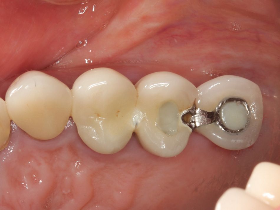
A 68-year-old female patient with unremarkable medical history presented in March 2013 with a failing maxillary implant prosthesis in the upper left posterior sextant. She reports that the upper left posterior sextant had become symptomatic over the last six months and believes that the TPS press fit implant was placed in the late 1990’s. Pressure to biting and swelling have convinced her to seek treatment although she has been advised of a deteriorating situation in the past.

Treatment planning

The patient understood that tooth #24, #25, and the implant in position #26 had a hopeless prognosis and would be lost. Her restorative options included either a fixed implant solution or removable prosthesis. However, the patient would only consider an implant supported fixed bridge.

The plan was to remove #24, #25, and #26 and regenerate the ridge prior to placing dental implants. Due to the size of the defect, and quantity and quality of bone which we would need to successfully place and restore the dental implants, it became clear that a two-stage procedure using a combination of Straumann® Emdogain® and Straumann® allograft was the treatment of choice.

Surgical procedure

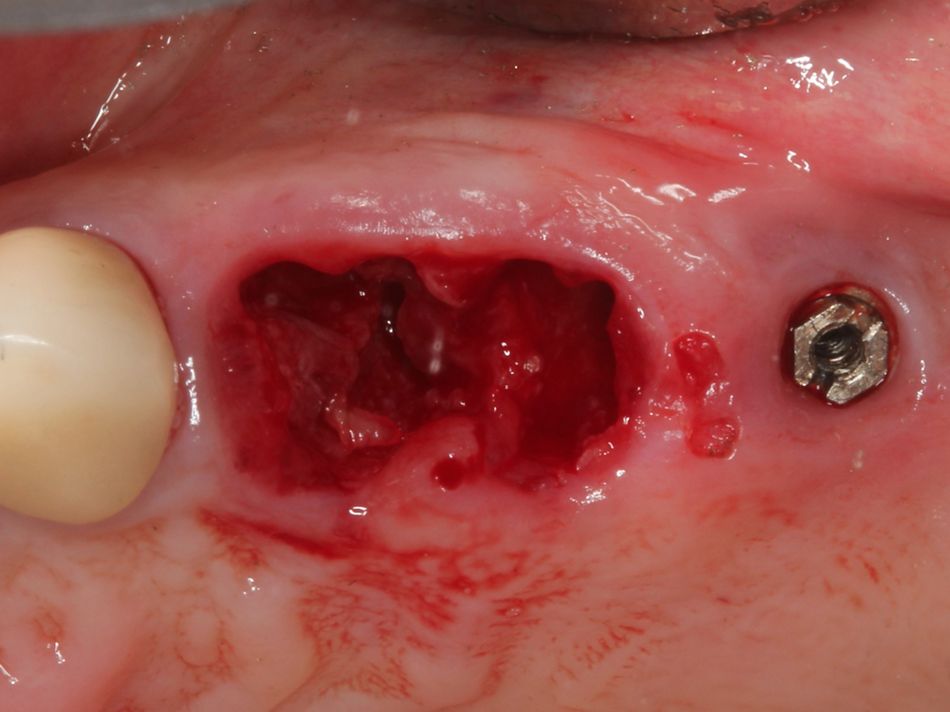
Standard surgical protocol was followed. The patient rinsed with chlorhexidine gluconate 0.12% prior to the surgery and was anesthetized with Lidocaine Hydrochloride 2%. After the restorations were removed, the teeth and implants were extracted with elevators and forceps. The area was thoroughly debrided of any soft tissue remnants and rinsed with chlorhexidine Hydrochloride 0.12%.

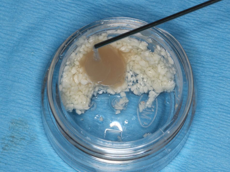
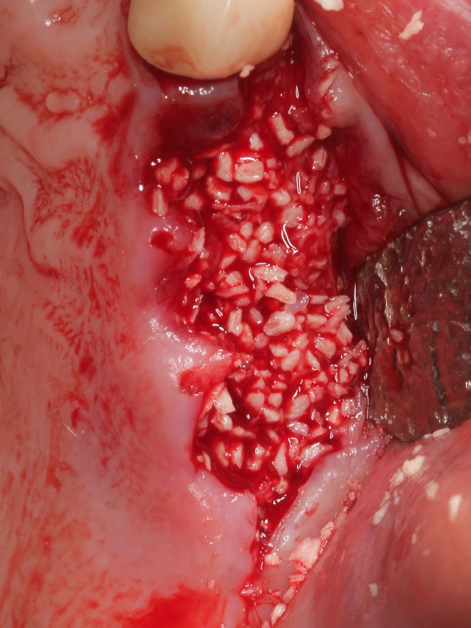
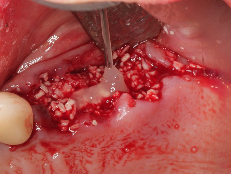
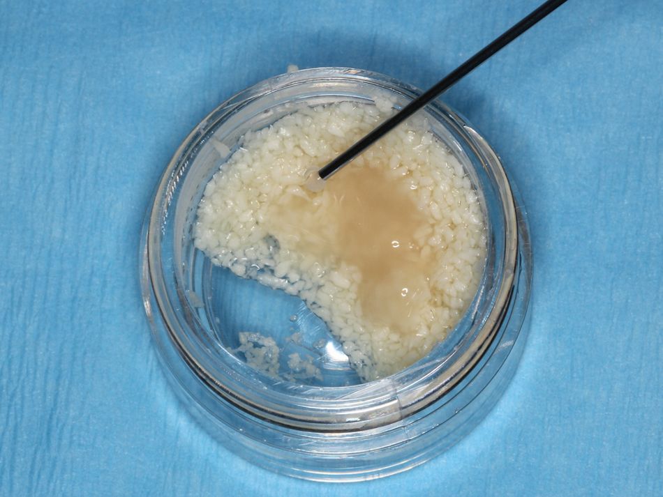
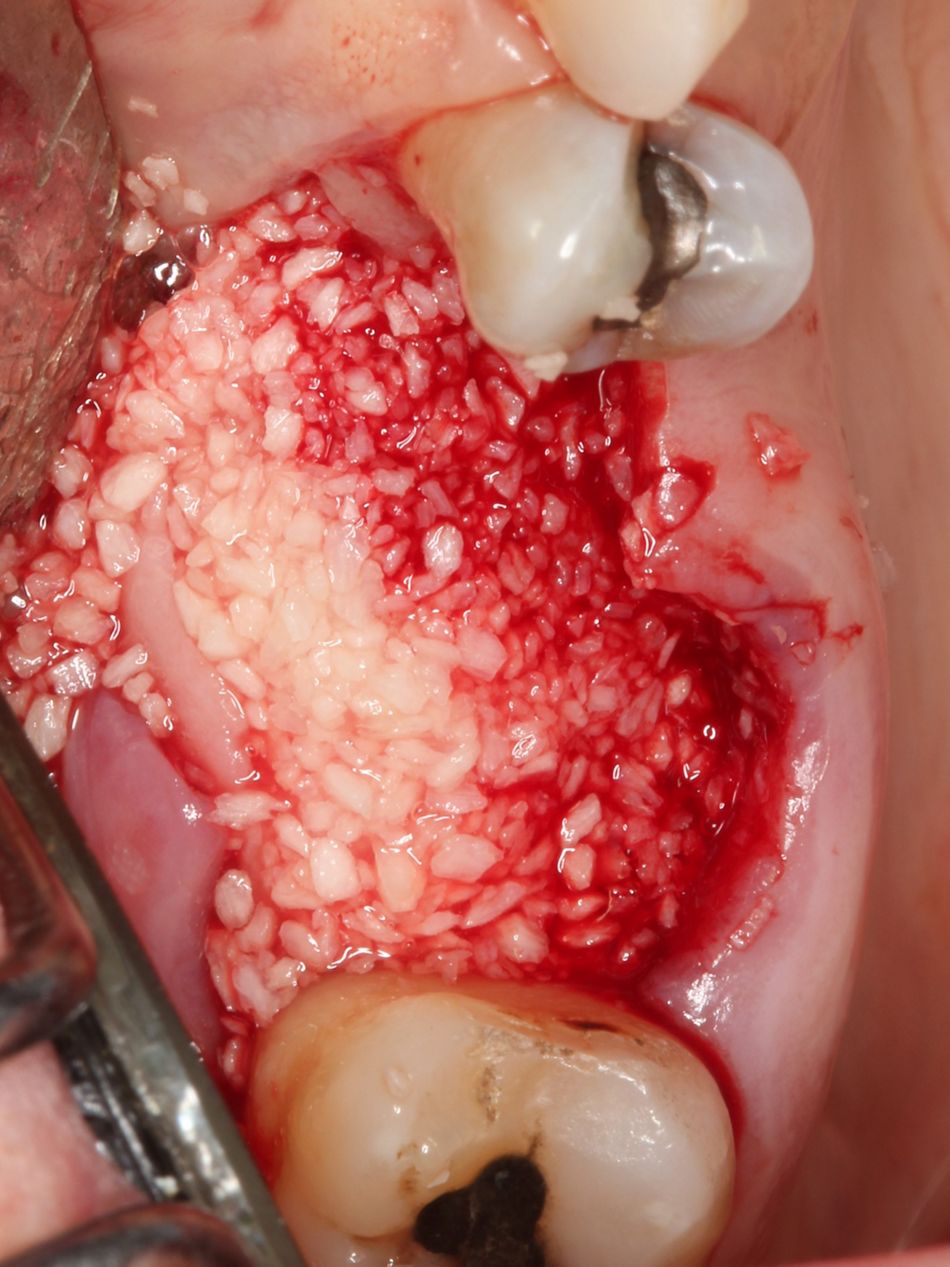
The decision to use Straumann® Allograft, 1.0 cc (250 – 1000 microns) - a mineralized freeze-dried bone allograft - was based on its resorption / turnover time and the fact that we had a 4-wall contained defect. The graft was hydrated with sterile water and mixed with Straumann® Emdogain® 0.15 ml prior to being packed in the defect. Straumann® Emdogain® was also placed at the suture line to aid in the soft tissue healing.

Treatment outcomes
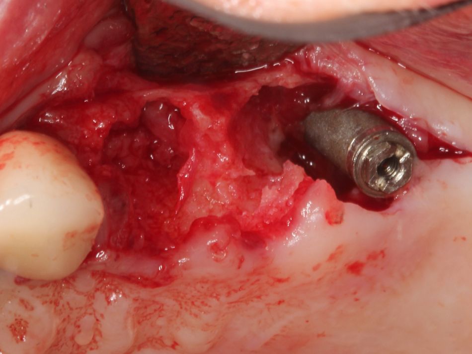
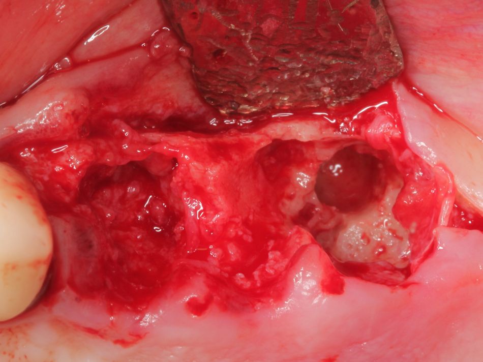
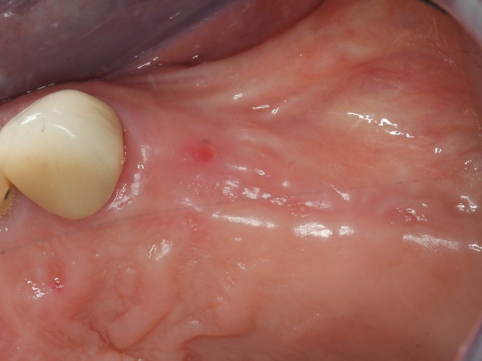
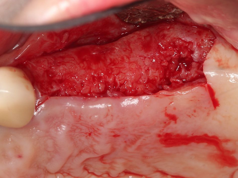
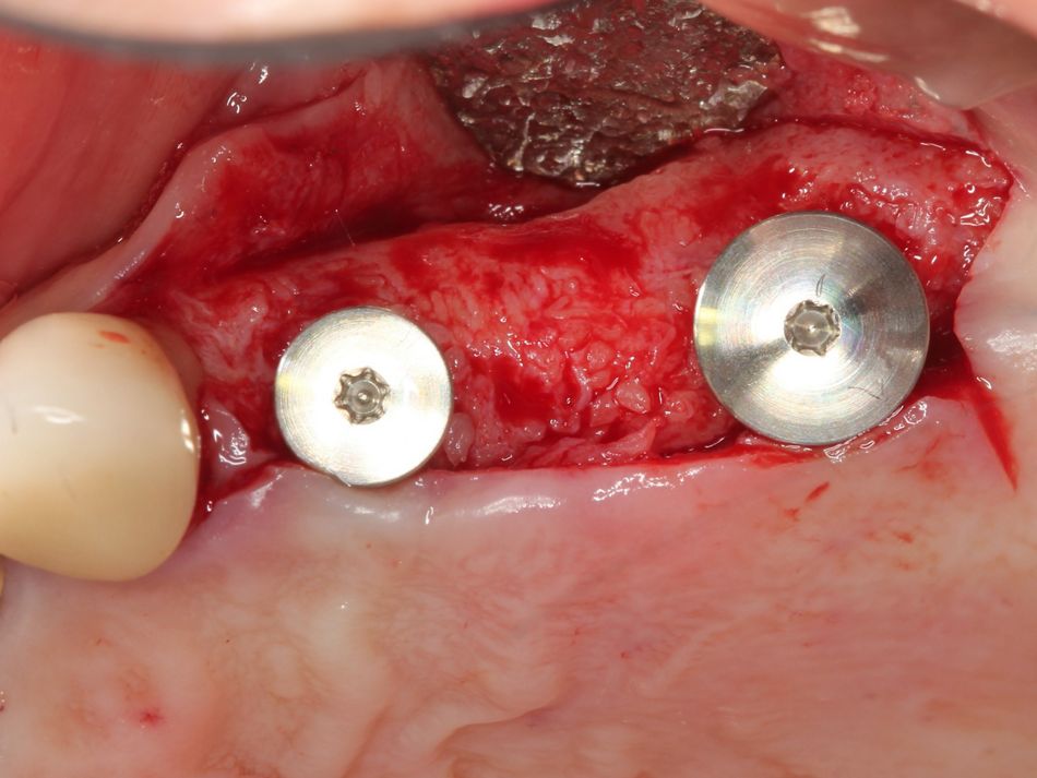
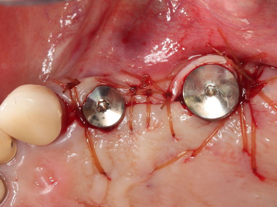
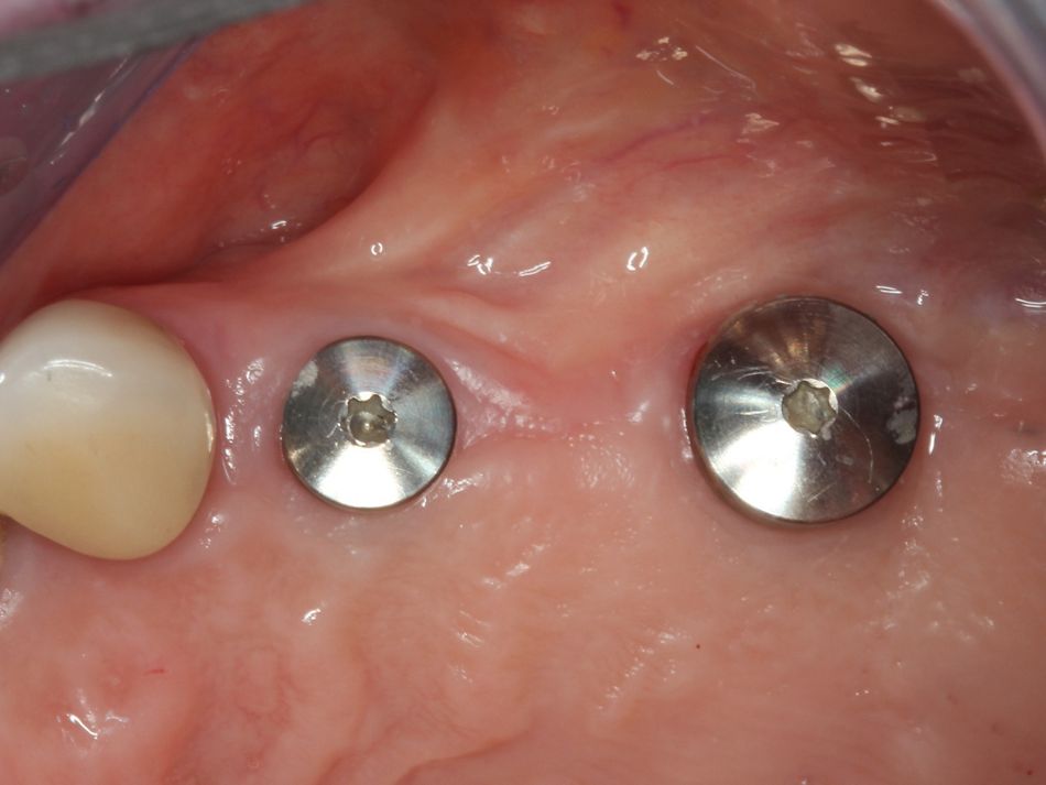
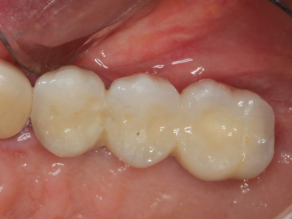
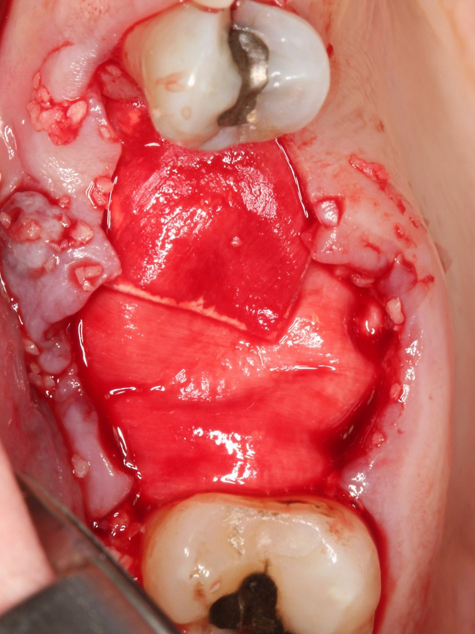
The area was reentered at four months reveling complete regeneration of the bone defects. This enabled the placement of a Straumann® Tissue Level Wide Neck SLActive® implant Ø6.5mm, 12 mm length in position #24 and a Straumann® Tissue Level Regular Neck SLActive® implant Ø4.8mm, 12 mm length implant was used in position #26. The fixtures were restored after ten weeks of hard and soft tissue maturation.

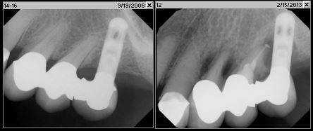
Follow up radiographs were exposed in 2014 and 2018 depicting the maintenance of a stable osseous crest. In 2019, 5 years after the final restoration, a stable result was registered as well.

Patient Nº2

Initial situation

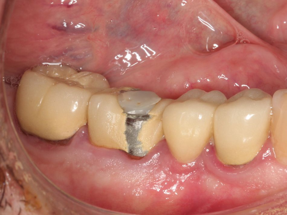
The patient, a 67-year-old male with unexceptional medical history, presents with a three-unit fixed bridge from #45 to #47. The distal abutment shows recurrent decay and possible root fracture. Excessive probing depths and drainage is associated with the defect.

Treatment planning

Treatment options include strategic extraction of tooth #47 with either four-unit fixed bridge or an implant solution with fixtures placed in position #46 and #47. The patient preferred dental implants as he was disappointed in the longevity of the previous tooth borne prosthesis. The patient was advised of the need for a two-stage procedure with implant placement after hard and soft tissue maturation following Guided Bone Regeneration.

Surgical procedure

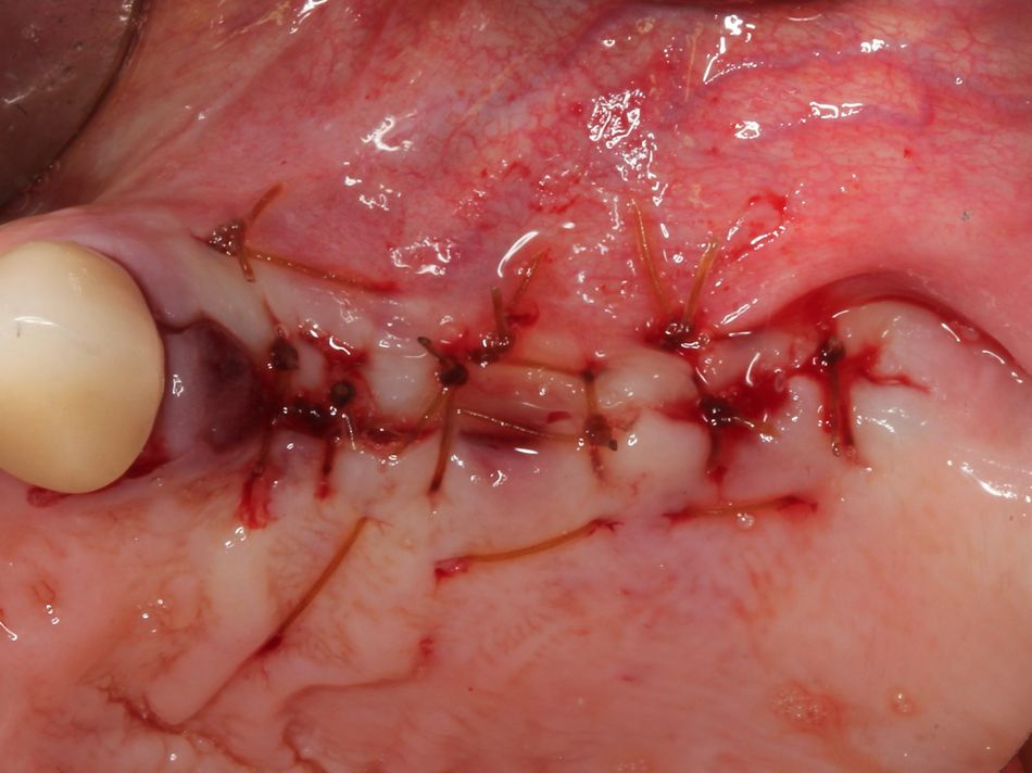
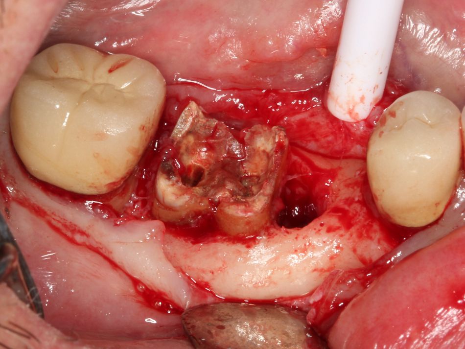
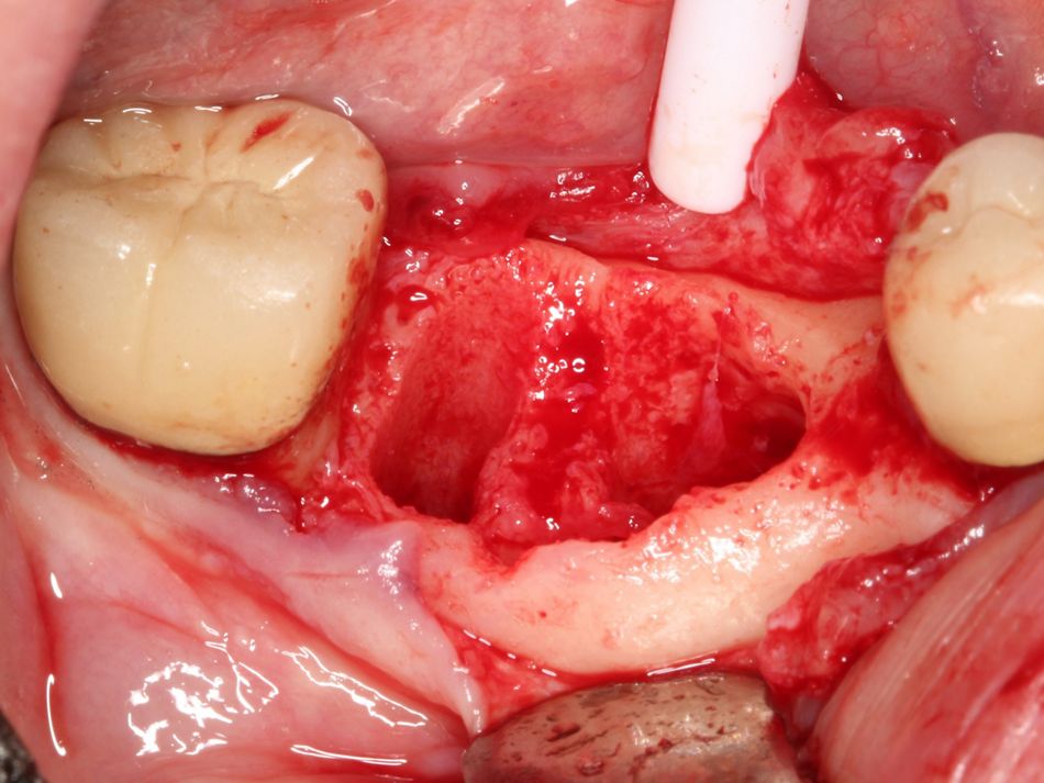
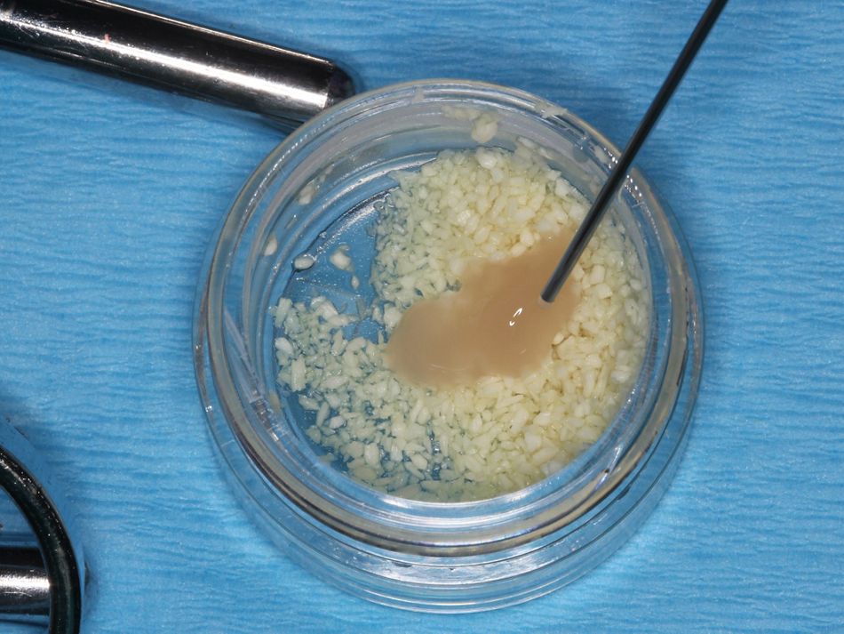
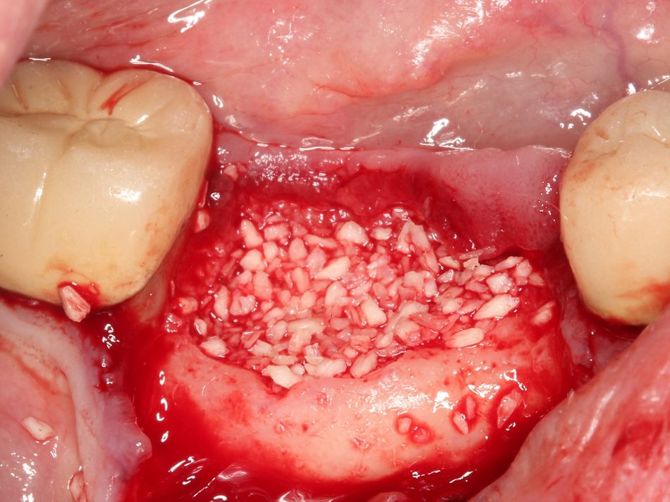
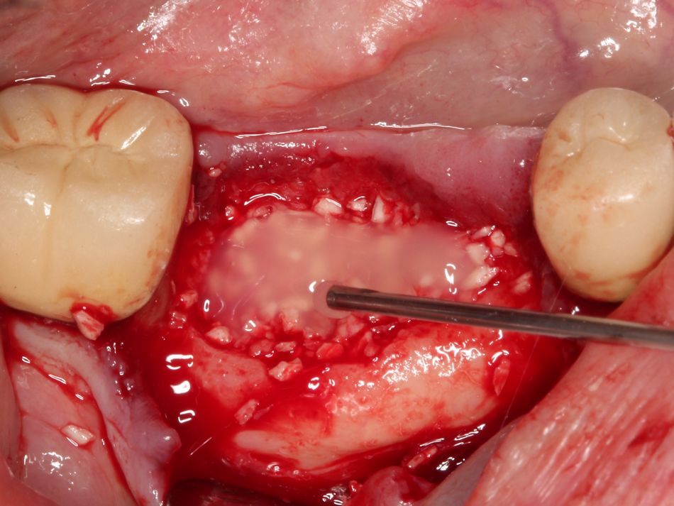
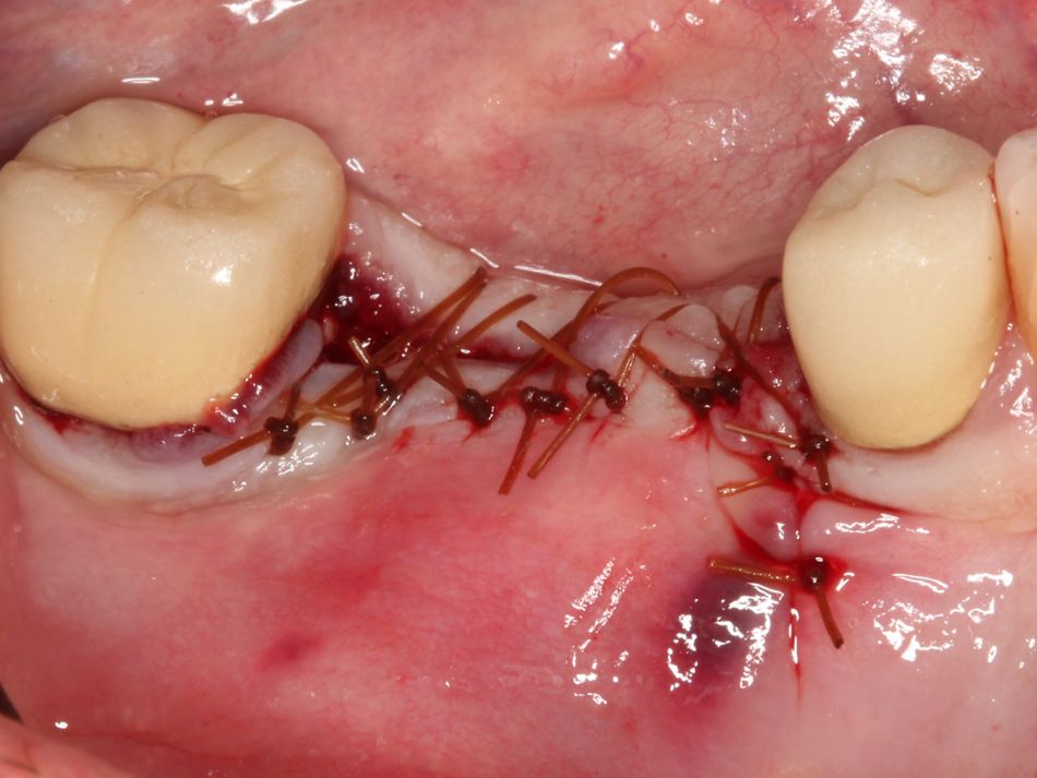
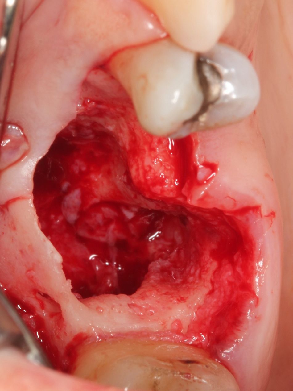
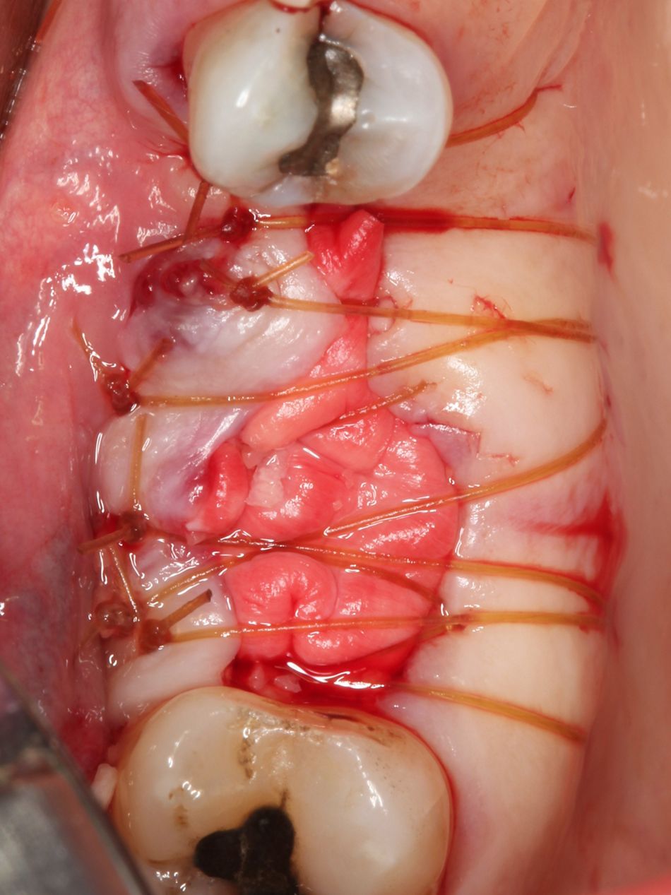
The bridge was sectioned distal to tooth #46 and the prosthesis was easily removed due to the excessive decay. Tooth #47 was extracted revealing a large defect which extended into position #46. Mineralized freeze-dried bone Straumann® Allograft, 1.0 cc (250-1000 microns) was hydrated and then saturated with Straumann® Emdogain® 0.15 ml. The defect was filled with the allograft material and then the remaining Straumann® Emdogain® was placed where we anticipated the superior aspect of the graft. Primary closure was attained with horizontal mattress suturing.

Treatment outcomes

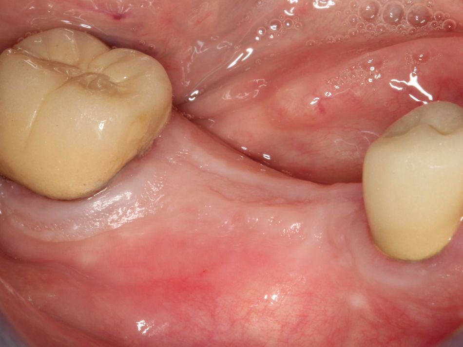
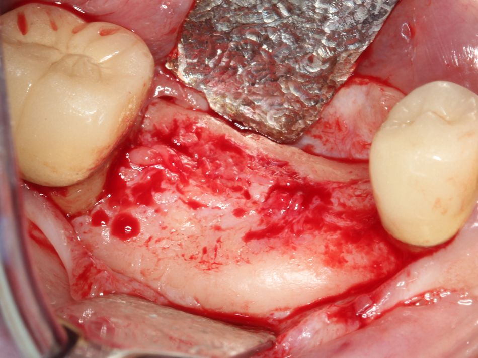
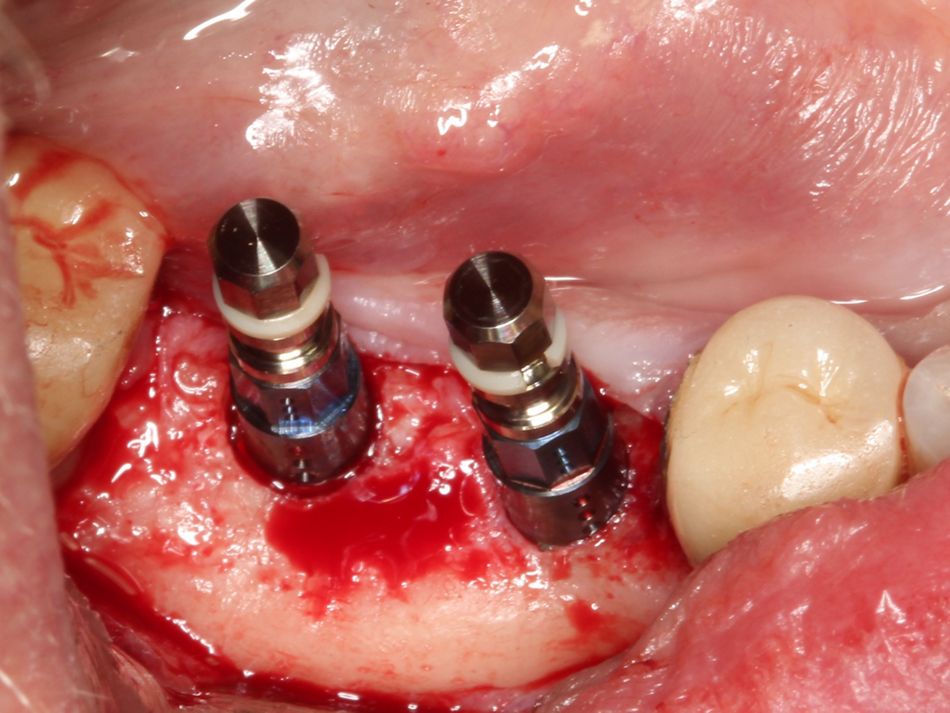
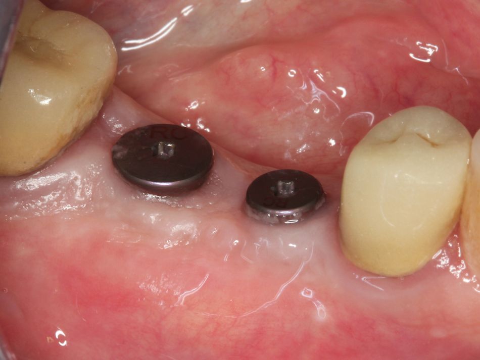
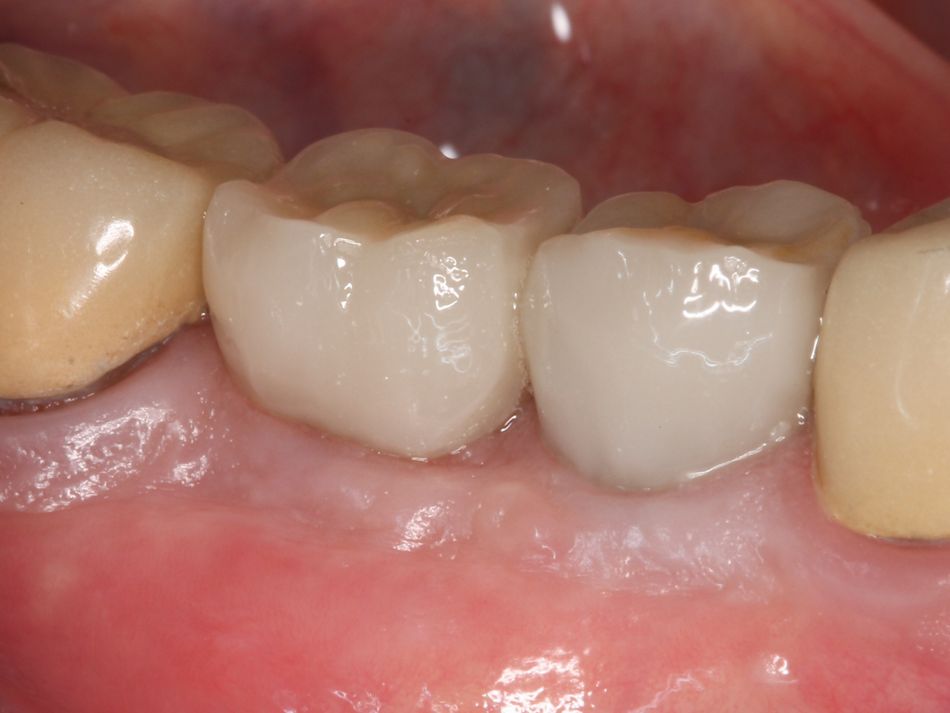
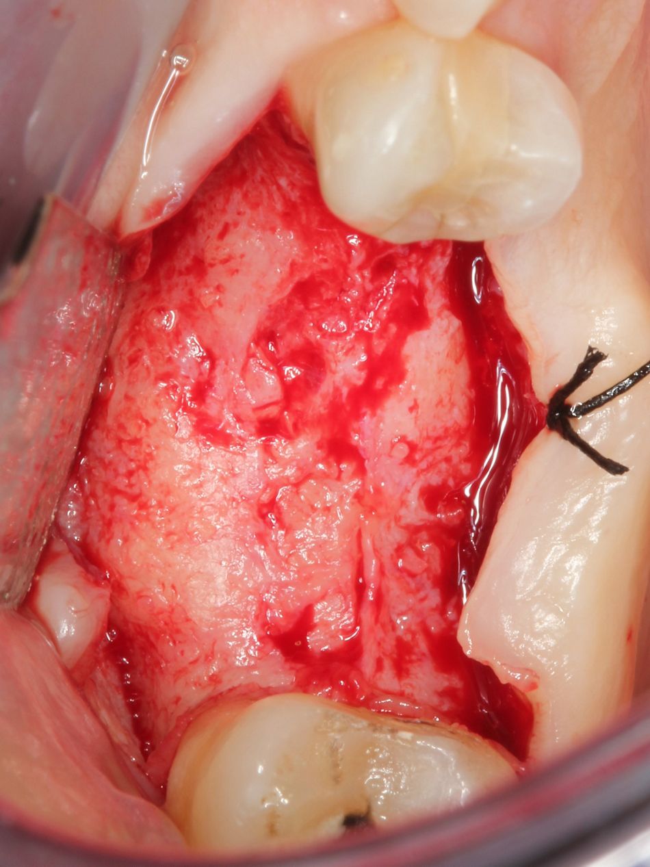
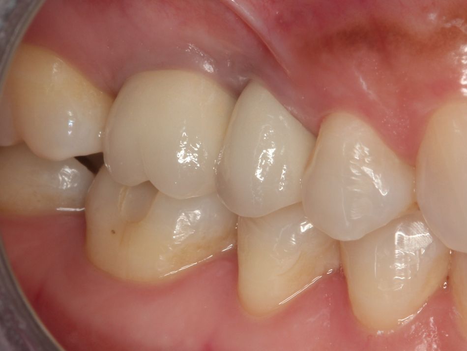
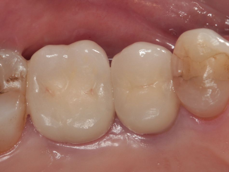
The area was opened after twelve weeks revealing complete regeneration of the defect. Two Straumann® Bone Level Regular Crossfit® SLActive® implants of Ø4.1 mm, 8 mm length and Ø4.8 mm, 10 mm length were placed in position #46 and #47. Healing was uneventful and ultimately restored as single units. Follow-up radiographs indicate a stable result after 5 years since the final restoration loaded the implants.

Patient Nº3

Initial situation

A 60-year-old female presented to our clinic in 2013. Her medical history indicates a non-insulin dependent diabetes. The patient presents with chronic infection around tooth #16. She was aware of an issue but has adapted. She elected to have treatment as the area had become symptomatic.

Treatment planning

The tooth was removed and revealed a very large defect that was debrided carefully in order not to damage the Schneiderian Membrane. The lesion would require GBR in order to place dental implants. Immediate placement was not an option as primary stability could not be attained because of sinus proximity.

Surgical procedure

After removal of any soft tissue remnants, freeze-dried allograft Straumann® Allograft, 1.0 cc (250 – 1000 microns) mixed with Straumann® Emdogain® 0.15 ml was placed in the extraction site. Primary closure could not be achieved, necessitating the placement of a collagen membrane.

Treatment outcomes

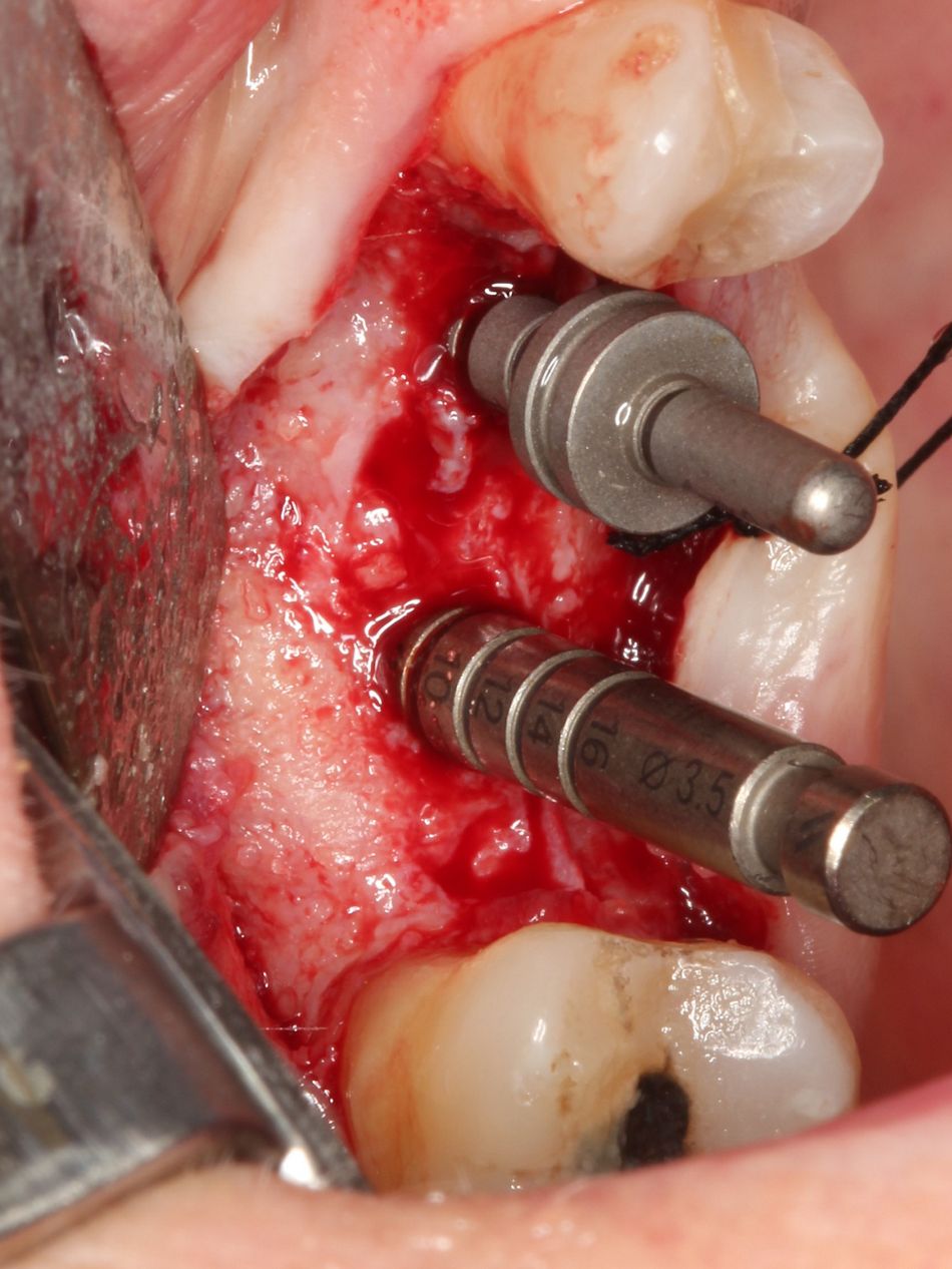
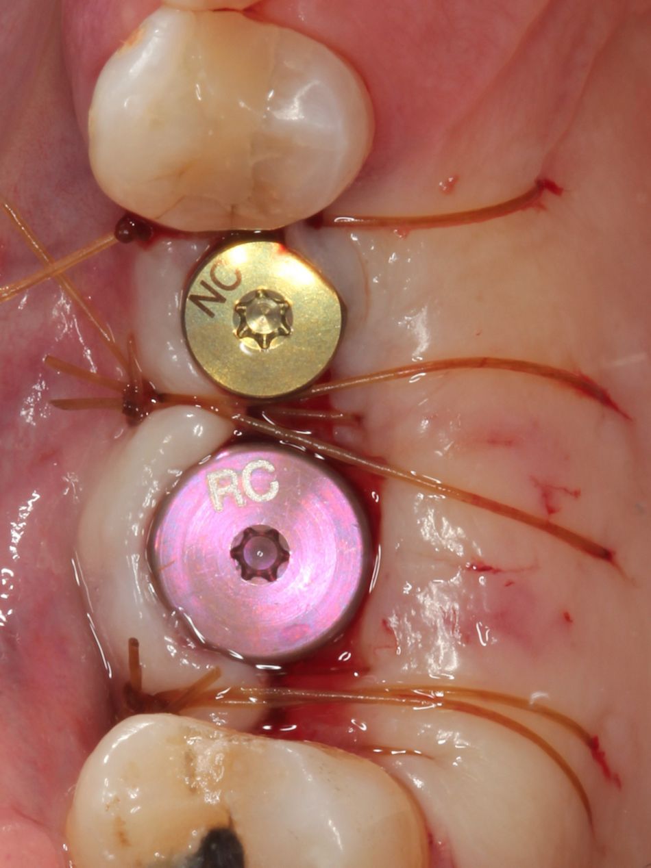
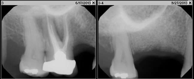
The site was opened after fourteen weeks of hard and soft tissue maturation revealing complete regeneration of the extraction site and giving us confidence that implants could be placed predictably. A Straumann® Bone Level Regular Crossfit® SLActive® implants Ø4.1mm, 8 mm length was placed in position #16 and a Straumann® Bone Level Narrow Connection 3.3 mm, 12 mm length was used in position #15, following the normal surgical protocol and allowed to osseointegrate. The fixtures were restored without incidence. The periapical radiographs indicated a stable bone level around implants at 2 and half and 5 years after the placement of the final restoration.