Introduction
Enamel matrix protein derivatives (EMDs) have been extensively evaluated in both pre-clinical (3) and clinical models. They are mainly composed of amelogenins, with smaller amounts of other non-amelogenin components, such as tuftelin, ameloblastin and enamel proteases (4). EMD is a biologically active compound that, once applied on a denuded root surface, starts a cascade of biological events, such as enhanced attraction and migration of mesenchymal cells, their attachment to the root surface (5), and differentiation into cementoblasts, PDL fibroblasts and osteoblasts. Enamel proteins enhance gene expression responsible for protein and mineralized tissue syntheses in PDL cells (6). This process may finally lead to reconstitution of the periodontal apparatus.
In this clinical case, we describe the successful regenerative therapy of a mandibular incisor with severe loss of periodontal support, utilizing Straumann® Emdogain® and a Connective Tissue Graft (CTG). This approach not only provided long-term stability for a tooth previously deemed to have a poor prognosis, but also helped meet the patient's expectations by allowing her to avoid complex surgical procedures and retain her tooth.
Initial situation
A 34-year-old female, healthy (ASA I) non-smoker with no medication, came to our practice complaining about mobility, gingival recession, bleeding, and suppuration from a mandibular incisor. The patient mentioned that the tooth began to move progressively several months ago, and her dentist applied a resin-bonded fixed splint between mandibular canines. Afterwards, she did not attend her follow-up appointments. The patient requested to keep her tooth.
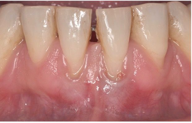
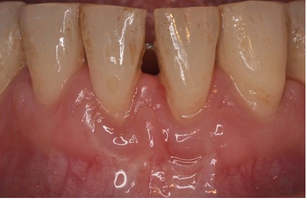
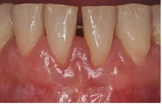
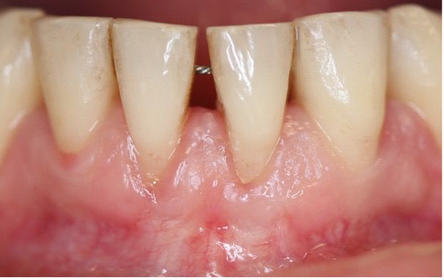
On the intraoral examination, a fixed splint was found from teeth #33 to #43; tooth #31 presented a thin gingival biotype, gingival recession on the buccal aspect with minimal attached and keratinized gingiva width, muscle and frenum pull, a probing depth greater than 6 mm, positive BOP (Bleeding on Probing), suppuration, redness and swelling, and dental plaque, suggesting a Stage III, grade B periodontitis (Fig. 1).
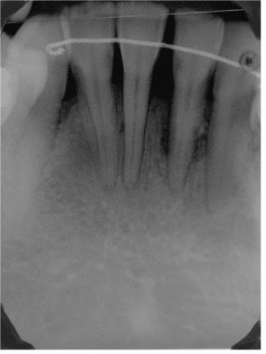
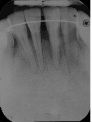
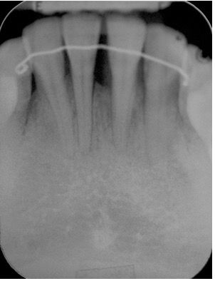
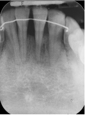
The radiographic examination revealed an extensive loss of periodontal support around tooth #31. (Fig. 2).

Treatment planning
- Cause-related non-surgical periodontal treatment with oral hygiene instruction and motivation.
- Periodontal reconstructive surgical treatment: Combination therapy with an autologous connective tissue graft and Straumann® Emdogain®.
- Supportive periodontal care every 3 - 4 months.
Surgical procedure
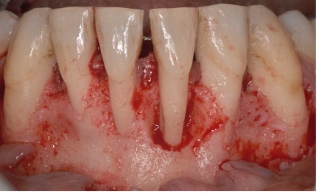
A full-thickness buccal flap was raised from teeth #33 to #43. After mechanical debridement of root surfaces and intrabony defects, an extensive 1-wall defect around tooth #31 was evident (Fig. 3).
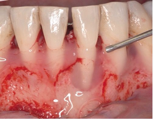
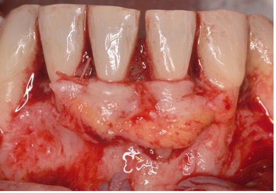
Following root surface conditioning with Straumann® Prefgel® for 2 minutes, Straumann® Emdogain® was applied onto denuded root surfaces and bony defects (Fig. 4).
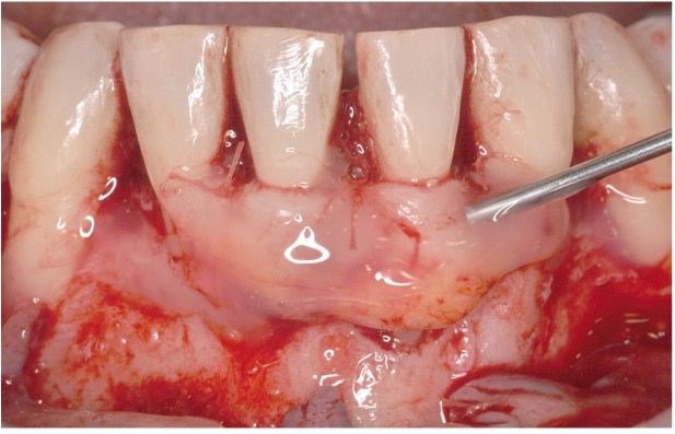
A connective tissue graft was retrieved from the patient’s palate and sutured to cover the denuded root surfaces (Fig. 5). Straumann® Emdogain® was also applied on top of the soft tissue graft (Fig. 6).
The single buccal flap was coronally displaced and sutured. Tenting sutures were also placed coronally to the contact area to stabilize the buccal tissues further.
Treatment outcomes
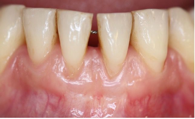
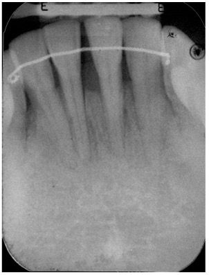
Clinical and radiographic outcomes were consistently satisfactory. At the 1-year postoperative evaluation, there was a minimal recession compared to the preoperative aspect, an increased attached and keratinized gingival width, with no frenum and muscle pull (Fig. 7). The radiographic 1-year control showed advanced hard tissue healing surrounding tooth #31 (Fig. 8).
At the 2-year postoperative evaluation, there was an improved gingival appearance, a certain degree of root coverage, together with an increased attached and keratinized gingival width (Fig. 9). The radiographic control showed stable results compared to the one-year situation (Fig. 10).
At the 5-year control, stable results over time were evident (Figs. 11,12).
The clinical evaluation at the 6-year postoperative control still showed stable results with minimal gingival recession (Fig. 13), and the radiographic evaluation revealed a complete resolution of the bony defect in tooth #31 (Fig. 14).
Author’s testimonial
Periodontal regeneration is a complex biological process that involves the de-novo formation of the lost tooth-supporting structures, including alveolar bone, periodontal ligament, and cementum over a previously diseased root surface. Combination therapies apply various treatment alternatives to improve outcomes further. The application of enamel matrix protein derivatives (Straumann® Emdogain®) on denuded root surfaces starts a cascade of biological events that may ultimately lead to the reconstitution of the periodontal apparatus.
In the presented clinical case, the outcomes showed probing depth reduction, clinical attachment gain with minimal gingival recession, increased attached and keratinizing gingival width, with no frenum and muscle pull together, and radiographic bone fill of the defects.