The Straumann® Bone Level Tapered Implant ⌀2.9 mm in daily clinical practice
A clinical case report by Kay Vietor, Germany
“The Straumann® Bone Level Tapered Implant ⌀2.9 mm represents an enrichment of my treatment portfolio that greatly simplifies the treatment procedures in cases with unfavorable narrow-space conditions often found in young patients.” Dr Kay Vietor
Author Kay Vietor

Kay Vietor
Dr. med. dent.
Private practice for oral surgery and implantology in Langen, Germany. Postgraduate education and degree in Oral Surgery. ITI-Fellow. Lecturer and author on CAD/CAM implant prosthetics and customized implant-borne prosthetic solutions, implant dentistry, 3D navigation in implantology, dental digital photography and digital archiving.
Related topics

Initial situation
The patient was a healthy, non-smoking, 19-year-old male, with dental agenesis of teeth 12 and 22 (Figs. 1,2). The patient underwent an orthodontic treatment due to a progeny tendency from 02.08.05 – 01.09.09 with a removable orthodontic appliance. Treatment with a fixed orthodontic appliance was performed from 29.11.2013 – 11.04.2016. After the orthodontic treatment was completed, appropriate gaps were made between the roots and the crowns of the adjacent teeth. The patient showed very limited gap widths and unfavorable interradicular space conditions. Initial marginal bone thickness in the gaps was approx. 4.5mm (Fig. 4) with with proclination of the apical base (Figs. 5,6).
Treatment planning
After discussing the situation with the patient, it was agreed that two small-diameter (2.9 mm) implants would be placed in positions 12 and 22, followed by fixed restoration. The position of the implants was carefully assessed using the CBCT technique (Figs. 5–8), and a 3D surgical drilling template was developed and printed to facilitate the drilling process (Figs. 9-12). Postoperative temporary prostheses were created to avoid gingival pressure and to secure the positional stability of the adjacent teeth (Fig. 3).
Surgical procedure
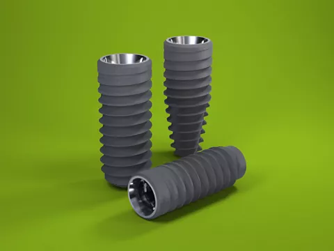
The implants were placed under local anesthesia, and the implant bed was prepared using a 3D-printed surgical drilling template. Two small-diameter Straumann implants (Bone Level Tapered, Roxolid®, SLActive® 2.9mm/12mm) were placed in positions 12 and 22 using the open flap technique (Figs. 13,14) followed by bone augmentation with autologous bone chips and a bone xenograft (Figs. 15,16). Jason® membranes and apical titanium pins were used to stabilize augmented regions. The respective protective caps were placed on the abutment part of each implant. The flap was repositioned and the wound closed with stress-free tight sutures (Fig. 17). The patient was prescribed antibiotic (2x 600mg clindamycin for 7 days) and anti-inflammatory (ibuprofen 400mg) therapy. The exact position of the implants was further checked and confirmed using the Ultra Low Dose CBCT technique (Figs. 18–20)
Prosthetic procedure
After 9 weeks of healing, a minimally invasive surgical procedure (puncture incision and displacement of the tissue) was performed in positions 12 and 22, and healing screws (SC H3.5) were placed (Figs. 21,22). An impression was then taken to produce temporary crowns for better gingiva formation and profiling (Fig. 23). Following the impression-taking (Figs. 24,25), individual restorations were designed and produced with Straumann® CARES® Visual software 10.1 (Figs. 26,27) and then placed in position.
Final result
Both the patient and practitioner were satisfied with both his extra-oral and intra-oral appearance after treatment. This was manifested by very good gingival development and also good pink/white esthetics (Figs. 28,29). Despite very limited gap widths and the unfavorable interradicular space conditions, the final result was very positive.