PURE Ceramic Implant Monotype (type 1A) in upper central incisor
A clinical case report by Alejandro Treviño Santos, Mexico, Alejandro Signorio Duque, Chile, Thomas Graber, Mexico
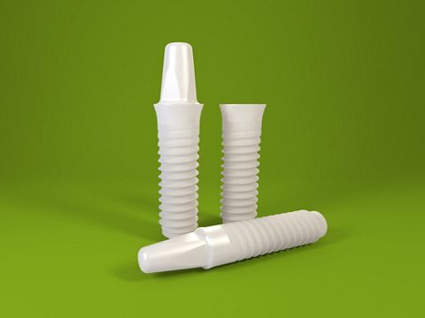
Patients’ esthetic expectations regarding dental implants have increased considerably, especially if an anterior tooth has to be replaced. Lately, more and more research has focused on ivory-colored implant materials to produce implants with improved esthetics but achieve high biocompatibility and the strength to withstand the forces present in the oral cavity at the same time.
About Alejandro Treviño Santos

Alejandro Treviño Santos
DDS, MSc
Chair, ITI Mexican Section
Member of the ITI Education Committee Group
Chair, ITI Scholarship Center, Mexico City
Associate Professor at the National Autonomous University of Mexico (UNAM), Postdoctoral and Research Division, Faculty of Dentistry, Mexico City
About Alejandro Signorio Duque

Alejandro Signorio Duque
DDS
ITI Scholar in Mexico City
Assistant Professor at San Sebastian University, Santiago, Chile
About Thomas Graber

Thomas Graber
SCDT
Master ceramist
Director of Estudio de Porcelana Suizo, Cuernavaca, Morelos, Mexico
Initial situation
A healthy 24-year-old male presented to the office with pain as well as second-degree mobility, shorter root size due to external resorption and a change in tooth color due to trauma to tooth 11. The patient wanted the restoration to look as if nothing had happened. (Figs. 1-2)
Treatment planning
We followed the workflow for Straumann® PURE Ceramic Implant Monotype, which comprises two procedures divided into four steps each. Step 1 of the surgical procedure was preoperative planning. This had to be prosthetic-driven to determine the most suitable position for the implant and prosthetic reconstruction. Step 2 was basic implant bed preparation, where a position indicator was used to ensure correct positioning of the implant and abutment. Step 3 would have been fine implant bed preparation to ensure that no excessive force was applied to the implant during insertion and tapping the thread into dense bone. However, we immediately placed an implant after extraction, so there was no need to perform step 3, and so our last step, which should have been step 4, was inserting the implant to the correct depth while making sure that the dots of the transfer piece faced buccally and lingually to reduce the risk of complications during the restorative phase. Normally, we begin the prosthetic procedure by placing a snap-on protective cap on the implant to keep its shoulder free of soft tissue during the healing phase, but in this case there was no need to do so since we had placed an immediate restoration. Therefore, step 1, which should have been step 2, was to place an interim prosthesis using an engaging temporary coping for at least 12 weeks. Accordingly, step 2 was to take a high-precision impression of the intraoral situation using a closed-tray impression technique so that we could perform step 3, the final step in this case, which was to create the final restoration and cement it in place.
Surgical procedure
Atraumatic extraction was performed (Figs. 3-4) resulting in a fresh socket with intact walls. (Fig. 5) The osteotomy was begun with a Ø 2.2 mm BL pilot drill to a depth of about 10 mm. Correct drilling was constantly checked since the design of the one-piece implant precludes any prosthetic readjustments. (Fig. 6) The position indicator was also used. (Fig. 7) A Straumann® PURE implant was placed with a primary stability of 32 Ncm. (Fig. 8) The gap between the buccal wall and the implant was filled with a slow-resorption xenograft. (Figs. 9-10)
Prosthetic procedure
Because the implant had excellent 3D positioning and good primary stability, a cement-retained interim prosthesis was built with perfect conformation of the emergence profile (Figs. 11-16) and no occlusion with the opposing tooth. (Fig. 17) During provisionalization a horizontal mattress suture was inserted to support the soft tissue and avoid any possible recession. (Fig. 18) Three months later a wide collar of keratinized tissue was visible with a great esthetic result. (Fig. 19) A closed-tray impression was taken and an analog was placed on the impression. (Figs. 20-21) The Straumann® PURE monotype implant must be restored with all-ceramic restorations, so it was decided to make five different ceramic copings to assess translucency: 1) an e.max LT A2, 2) an e.max HT A2, 3) a Sagemax NexxZr Trans, 4) a ZirCAD MO, and 5) a ZirCAD Super Trans. (Fig. 22) If translucency was needed for the restoration an e.max HT coping (lithium disilicate) was the best option, but if translucency needed to be blocked a ZirCAD coping (zirconia) was the most suitable solution. (Fig. 23) To optimize the visual effect of the translucency of the copings, we made a zirconia analog (Figs. 24-25) to test the translucency of the ceramic copings and assess how they would behave when cemented in place.
Treatment outcome
The chief complaint of the patient was addressed and his expectations surpassed. (Figs. 26-27) It is vital to remember that the implant is designed to have the implant shoulder sit 1.8 mm above the crestal bone. If we follow this basic principle it is fairly simple to adhere to the recommendations of the 5th International Team for Implantology (ITI) Consensus Conference regarding the clinical performance of screw- versus cement-retained implant-supported fixed reconstructions, where the evidence shows that we must avoid a cement margin and thus the possibility of cement residue (prosthetic margin ≥ 1.5 mm supragingivally). It is important for the clinician to understand that the procedures involved with cement retention for implant-supported crowns are not simple and should be carried out with caution. The selection of the implant placement and loading protocol can have a negative impact on survival and success if specific criteria are not adhered to and/or the clinical procedure is not executed to a high enough standard. The benefits of the type 1A placement and loading protocol for the patient (6th ITI Consensus Conference) should be carefully considered, as should the associated risks.
Findings
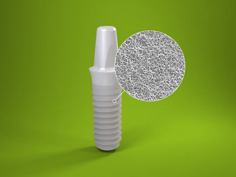
The Straumann® PURE monotype implant is coated with the ZLA® surface whose macro- and micro-roughness is designed to provide a structure for cell attachment equivalent to SLA®-treated titanium as regards osseointegration, peri-implant bone density, and bone-to-implant contact ratio. This makes it feasible in immediate implant placement. The one-piece design of the implant means that the abutment is built into the implant to eliminate any microgap between the implant and the abutment. This provides great support for the peri-implant soft tissues, thereby creating an emergence profile that allows the implant restoration to mimic the soft tissue of the contra-lateral tooth.