Predictable horizontal ridge augmentation and soft tissue thickening using an allogenic bone strut and a porcine collagen membrane
A clinical case report by Sebastian Stavar, Netherlands
Various reports have demonstrated the superiority of implant supported dental restorations over removable prosthetics in terms of patient acceptance, aesthetic appearance and quality of life.1 The prerequisite for successful and stable implantation of a fixed restoration is a sufficient amount of residual bone in which the implant can be submerged. Edentulism, however, may significantly promote bone resorption and, thus, compromise the feasibility of placing a fixed implant. While a smaller implant diameter and the introduction of short implants represent excellent implant-based approaches to overcome limitations in the lack of both horizontal and vertical bone height, extensive bone atrophy may not allow these systems to be implanted. Despite the advantages of implant supported restorations, prosthodontists tend to advise a shift in treatment to removable prosthetics in these situations, whereby the patient’s desire for permanent rehabilitation remains unmet. To address this issue, several methods for alveolar bone augmentation, predominantly based on the harvesting of autologous bone tissue, have been developed.2 While these procedures represent valuable solution for predictable and efficient bone augmentation, bone substitute materials, especially allogenic bone grafts, have the potential to dispense with the need for bone harvesting.3, 4
Initial situation
A 66-year old female, non-smoker in good general health, presented with missing teeth in sites 36, 45 and 46, which were extracted due to caries and fracturing. While the missing teeth in sites 36, 45 and 46 were initially restored with bridges, the patient wished for a fixed implant-supported prosthetic restoration in sites 45 and 46, due to a failure of the bridge. However, a pronounced loss of bone tissue in the posterior mandible (Cawood and Howell Class III), especially concerning the buccal wall, was observable at the initial assessment and further confirmed by radiographic imaging, so that direct implant placement was impossible (Figs. 1-3).5 The thin soft tissue in the region concerned posed an additional problem for proper treatment and aesthetic rehabilitation (Fig. 2).
Treatment planning
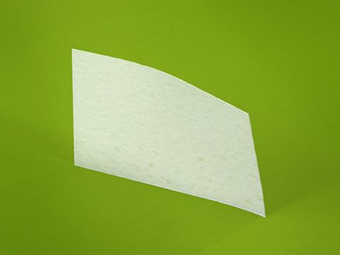
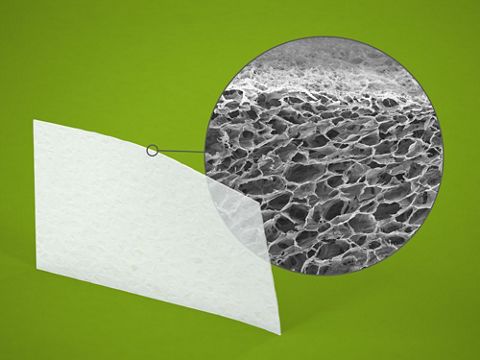
Several treatment options for restoring the lost bone and soft tissue were discussed with the patient in order to facilitate the placement of fixed dental prostheses and provide a positive overall aesthetic outcome. As the patient refused the option of harvesting autologous bone and soft tissue, a staged guided bone and tissue regeneration procedure using allogenic bone grafts and porcine collagen membranes was determined to be the best, and simultaneously least invasive, course of treatment meeting the patient’s requests. The treatment plan included the restoration of the horizontal mandibular bone defect with an allogenic cortical strut (maxgraft® cortico) and allogenic granules (maxgraft® cancellous granules), followed by soft tissue thickening using a porcine dermal collagen matrix (mucoderm®) together with the placement of two Straumann Roxolid® SLActive® BLT implants with a diameter of 4.1 mm and length of 12 mm.
Surgical procedure
The horizontal augmentation of the mandible was conducted using the modified shell technique.6 A crestal incision in the gingival margin followed by mesial and distal relief incisions were made to expose the defect (Fig. 3). The periosteum was carefully separated from the host bone on both the lingual and buccal sides in order to achieve proper soft tissue mobilization. Using a piezoelectric device, the cortical layer of the residual bone was perforated in order to create a bleeding recipient site, which accelerates the vascularization and vitalization of the allogenic grafting material (Fig. 4).7 The allogenic cortical strut was shaped with a diamond burr to match the defect and attached to the mandibular bone using titanium osteosynthesis screws (Fig. 5). As the cortical strut remains avital and implants need to be placed in vital bone to ensure proper osseointegration, the space between the cortical strut and the residual bone was filled with cancellous allogenic bone chips rehydrated in blood, which will eventually be remodeled into vital bone tissue (Figs. 6,-7).
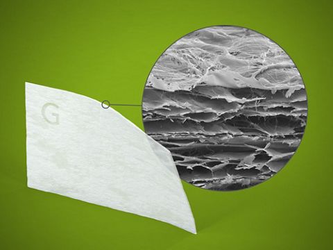
Prosthetic procedureAfter the edges of the cortical strut were blunted, the surgical site was covered with a pericardium collagen membrane (Jason® membrane), which was adapted to the defect and fixed onto the ridge using titanium pins (Fig. 8). The fixation of the membrane is important, as it prevents both the displacement of the grafting material and irritation of the mandibular nerve (mental nerve). In order to prevent soft tissue perforation, an additional membrane was placed over the augmented ridge, and a combination of mattress and single-button sutures was used to maintain proper, tension-free wound closure (Fig. 9). A panoramic radiograph and two CBCT-scans were recorded to check the situation before and after the intervention in order to demonstrate the pronounced horizontal bone augmentation and assess changes in radiopacity and bone volume (Figs. 10-12).
Following 5 months of uneventful healing, the patient presented for the next procedure, which included the insertion of two fixed dental implants and simultaneous soft tissue thickening (Fig. 13). The previously placed incision lines were reopened in order to remove the titanium osteosynthesis screws used for cortical strut fixation (Fig. 14). Vital, bleeding and robust bone was found within the granule-filled space adjacent to the avital cortical strut, which was flush-mounted in the new formed bone tissue (Fig. 15). Horizontal bone gain was sufficient for stable and fully submerged implantation of the designated Straumann BLT implants with a torque value of 35 Ncm (Figs. 16-17). Subsequently, cover screws were inserted in the implants, and bone substance, which was being removed during placement of the pilot drill, was used for contouring around the implant shoulders (Fig. 18). Prior to wound closure, which was performed with double-sling sutures, a porcine collagen matrix (botiss mucoderm®) was placed over the augmentation site to thicken the soft tissue (Figs. 19-20). A radiographic control image was recorded in order to assess implant position and bone density within the augmented area (Fig. 21).
Following 3 months of healing, the thickened soft tissue presented a natural and healthy appearance, so that papilla shaping could be achieved by the installation of gingival formers to set the course for a positive aesthetic final outcome (Figs. 22-23). Well-conditioned soft tissue was found 5 weeks later when the gingiva formers were removed and the final prosthetic restoration was installed (Fig. 24).
Prosthetic procedure
Loading of the implants in sites 45 and 46 was performed using a chair side implant workflow. Therefore, a Ti-base (Straumann® Variobase® C) was used for intraoral scanning (Omicam, Cerec, Sirona) and the subsequent production of CAD/CAM fabricated (Cerec, Sirona) screw -retained hybrid- abutment- crowns. The individually customized abutments promote the ideal emergence profile of the gingival margins. After crystallization, staining and glazing, the final crowns perfectly matched the coloration of the patient’s dentition and supplied a natural and aesthetic appearance, as well as a tight and functional occlusion (Figs. 25-26). Radiographic check-up at the time of implant loading again confirmed the excellent bone gain and the absence of resorption within the augmented area (Fig. 27).
Final result
The entire treatment course was conducted in accordance with the initial planning and resulted in an optimal final outcome (Figs. 28-29). The dental restoration in sites 45 and 46 with two implant-supported prostheses exactly met the patient’s request, and the result fulfilled the overall expectations. In addition, the soft tissue thickening procedure generated a notable increase in soft tissue thickness, with healthy coloration, and significantly enhanced the aesthetic appearance. The fixed implants remained completely osseointegrated 18 months after implantation, and not even the slightest reduction in radiopacity was observable, demonstrating an entirely successful outcome of the planned treatment (Fig. 30).
Conclusion
The patient had requested the installation of fixed dental prostheses. Extensive bone and soft tissue augmentation was necessary, but the patient decided against harvesting of autologous tissue. The treatment plan demonstrated here, using bone and soft tissue substitutes, was completely in accordance with the patient’s desires. The modified shell technique conducted with an allogenic cortical strut and cancellous allogenic granules resulted in significant horizontal bone gain, and facilitated the prosthetic restoration requested by the patient. It is worth noting that the observed results at the follow-up visit exactly conformed to the expectations of the planned surgery, which underscores the high predictability of the guided bone regeneration technique employed here. To nsure optimal implant stability, the Straumann® BLT SLActive® implant was chosen, as its surface demonstrated 60 % more bone-implant contact two weeks after insertion compared to the standard SLA® surface, and additionally allows for completely submerged implantation and, so does not impair flap closure.8 The soft tissue augmentation procedure conducted with a porcine collagen matrix significantly increased soft tissue thickness and simultaneously saved the patient the need for additional surgical sites for tissue harvesting. The utilized implants remained well integrated in the augmented bone 18 months after implantation, and a positive aesthetic final outcome was achieved. Overall, this case demonstrates that, while being far less invasive, the clinical performance of the modified shell technique with allogenic bone grafts is comparable with the conventional shell technique established by Prof. Dr. Khoury.


