Rehabilitation of the edentulous maxilla with four immediately loaded implants
A clinical case report by Mehmet Akif Eskan, Turkey
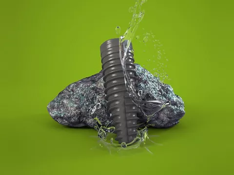
This case report shows how a hopeless maxillary dentition can be restored with an immediately loaded functional prosthesis in a patient with poor bone quality and demanding esthetic and functional requirements. Straumann® Bone Level Tapered (BLT) implants and Straumann® Screw-Retained Abutments (SRAs) provide the necessary implants and prosthetics to support this Straumann® Pro Arch solution.
Author: Mehmet Akif Eskan

Mehmet Akif ESKAN
D.D.S., PhD
Diplomate of the American Board of Periodontology. Adjunct Faculty, Department of Periodontics NOVA Southeastern University College of Dental Medicine, Fort Lauderdale, Florida/USA. Private Practice in Istanbul.

Initial situation
A 42-year-old male patient in good physical health presented at our clinic with the chief complaint of loose teeth and poor esthetics (Fig. 1). He reported that he was a former smoker and did not take any medication. His oral hygiene was poor, with a plaque score of 90% and bleeding on probing of 86%. Following the clinical and radiographic examination, it was determined that his remaining upper teeth and teeth 31, 41, 44 and 47 had a hopeless prognosis (Figs. 2, 3). Furthermore, CBCT analysis revealed a rounded and dome-like lesion in the left maxillary sinus (Fig. 4). After a consultation with an ENT specialist, this was diagnosed as a retention cyst of the maxillary sinus, which can regress spontaneously and disappear in 17 to 38 percent of cases [1].
Treatment planning
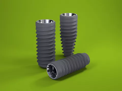
The treatment for the upper jaw included the extraction of hopeless teeth. Furthermore, since the patient had never previously tolerated a removable prosthesis, he requested a fixed restoration to restore his dentition during the treatment period. In order to meet the patient’s expectations and shorten the treatment time, the insertion of a screw-retained provisional prosthesis immediately following the extractions and implant placement were planned. Four implants (Straumann® BLT, Ø 4.1 mm, Roxolid, SLActive® implants), with tilting of the two posterior implants, were planned for placement. As the goal was to load the implants immediately, it was critical for primary stability to be established for immediate functioning with the planned fixed prosthesis.
Surgical procedure
Surgery was carried out under local anesthesia. All upper teeth were extracted, and a full thickness flap was reflected to enable all the granulation tissue to be completely removed (Fig. 5). A clear acrylic surgical guide was made to help us visualize whether at least 8mm of interocclusal space was available (Fig. 6). A small window to the sinus was opened with a round bur to locate the anterior sinus wall (Fig. 7). Where necessary, countersinking was done to secure both buccal and lingual cortical bone contact at the implant collar in the thin bone crest. The posterior implants were placed with about 45°-30° of inclination, and 30° angulated SRAs were placed on the posterior implants to correct the angulation for immediate prosthetic rehabilitation (Fig. 8). Following placement of the SRAs, a provisional full-arch acrylic prosthesis using temporary titanium copings was delivered on the day of surgery. The provisional prosthesis was screwed into the patient’s mouth, and the esthetics and occlusion were checked (Figs. 9-11). The patient was instructed to maintain oral hygiene with chlorhexidine 0.2% mouthwash twice a day for the first week post-surgery, followed by a water flosser (Waterpik®). The patient was also instructed to maintain a soft diet for 6 weeks post-operatively.
Prosthetic procedure
After four months of the healing, the provisional prosthesis was removed (Fig. 12). The implants and peri-implant soft tissues showed good stability and no signs of mucositis or peri-implantitis after clinical and radiographic examination. Open-tray impression posts for screw-retained abutments were attached to all implants, and were splinted using pattern resin and wires to avoid any unpredictable distortion (Figs. 13, 14). An open-tray impression was taken using a dimensionally stable polyether siloxane material (Fig. 15). Furthermore, a plaster model was cast, and the provisional prosthesis was seated on the model to check the adequacy of the impression. As the patient was happy with the esthetics of the provisional prosthesis, it was scanned for construction of the CAD/CAM framework (Figs. 16, 17). Following all try-ins, including metal and porcelain options, the prosthesis was delivered (Figs. 18, 19).
Treatment Outcome
The short edentulous span in the lower anterior region was restored with an implant-supported fixed bridge, and the 44 gap restored with a single implant-supported crown. This new implant design allows immediate implant placement and functional loading. The final x-ray showed the prosthesis precisely fitted on the SRAs (Fig. 20). At 31 months after loading, the implant survival and success rate was 100%, with no mechanical or biological complications. The overall functional and esthetic outcome of the treatment was judged to be excellent by the patient and the clinician (Fig. 21).