Application of the Straumann® BLT Ø 2,9 mm implant in the esthetic zone
A clinical case report by Wiebe Derksen, Netherlands
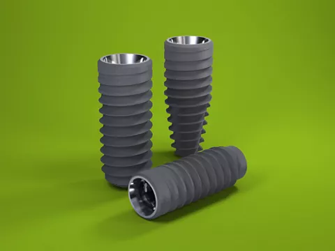
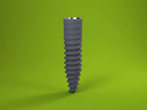
The restoration design described in this case report guarantees a biocompatible transmucosal zone and prevents cement excess. Bone remodeling is reduced thanks to the asymmetrical Variobase® abutment and this biocompatible restoration design (Fig. 19). The new implant diameter gives us clinicians the opportunity to provide our patients with clinically acceptable implant restorations even in confined spaces (Fig. 20).
Author: Wiebe D.C. Derksen

Wiebe D.C. Derksen
Graduated Cum Laude in dentistry from the University of Groningen in 2010. In 2012 he started his PhD at ACTA (Academic Center of Dentistry Amsterdam) on digitization in implant dentistry. In 2015 he graduated from the ACTA 3-year “Oral Implantology and Implant Prosthodontics” postgraduate program. He is a co-author of the current 5th ITI Consensus Review on Computer Guided Surgery, the chairman of the Dutch Society of Young Implantologists, the editor of the Quality Practice dental journal, was awarded an ITI clinical research grant in 2013 and became an ITI Fellow in 2015. His current expertise and research interests focus on guided surgery, intra-oral scanning, abutment choices, CADCAM and chairside (implant) restorations.
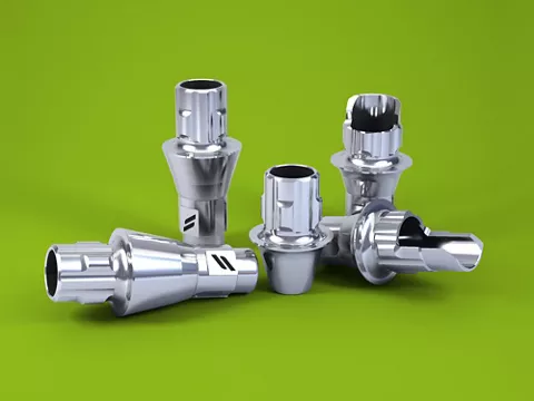
Related products
Initial situation
A 21-year-old male patient presented with a missing tooth 31 after an earlier trauma. The clinical diastema was only 4.5mm, whereas 5.5mm of space was available at bone level (Figs. 1,2).
Treatment planning
The CT image showed sufficient bone width for implant placement without prior bone augmentation (Fig. 3). However, since the anatomy of the underlying bone would direct the implant into a buccal angulation, a buccal screw access hole or cement-retained restoration was therefore inevitable. Figure 4 demonstrates the clinical situation directly before implant surgery.
Surgical procedure
After the administration of local anesthetics, a small midcrestal incision – without releasing incisions – was made. The osteotomy was prepared using the new 1.6mm needle drill and finished using the 2.2mm pilot drill (Figs. 5,6). Since the bone was soft, no profile drill or tap was employed. The osteotomy was created within the bony envelope, and the future implant site was surrounded by native bone, although a slight buccal contour change was discernible (Fig. 7). A Straumann® BLT SC 2.9mm/12mm SLActive® implant was selected and inserted in the correct buccal orientation (Figs. 8-10). Although the implant was surrounded by bone, due to the contour dip it was decided to augment the buccal contour (Fig. 11). In view of the shape of the lower incisors and the limited space, a special asymmetrical two-piece healing abutment was designed by the manufacturer and placed accordingly (Figs. 12,13). After abutment insertion the buccal contour was augmented using a slowly resorbing DBBM-graft (Fig. 14). Two single interrupted sutures (polypropylene 6-0) were used to close the surgical site. Directly after surgery a periapical radiograph was obtained to verify the implant position (Fig. 15).
Prosthetic procedure
Three months after implant placement the implant was well osseointegrated, and an open-tray conventional impression was taken (Fig. 16). Due to the buccal angulation of the implant it was decided to create a screw-retained zirconia coping luted on a 2mm Variobase® abutment with an adhesively cemented lithium disilicate veneer (Figs. 17,18).