Post extraction implant placement with bone and soft tissue graft combined with immediate provisionalization in a damaged socket (BLT/Variobase)
A clinical case report by José Manuel Losada, Spain
This case shows that, while it is necessary to continue to deepen and analyze this procedure in the long term, the results obtained to date are equivalent to, or more favorable than, those achieved when procedures are performed in stages. These results have been very motivating to continue to deepen this therapeutic strategy. The procedure offers the main advantages of exposing the patient to fewer surgical procedures, a reduction in maneuvers and prosthetic sessions, with a consequent decrease in clinical time and total treatment time. This technique requires extensive training as it is very technique sensitive due to the importance of the 3D implant position, the volume of soft and hard tissues and the prosthesis design.
Initial situation
The 28-year-old male patient came to our practice with no general health conditions or habits that could affect the prognosis of our treatment. After clinical examination (Fig. 1) (periapical x ray, probing, CBCT scan), a vertical root fracture was diagnosed, accompanied by a defect on the buccal and palatal walls. Various treatment options were presented and discussed together with the patient, and he decided to have tooth 21 replaced with an implant. As his oral hygiene was poor, basic periodontal therapy was performed and plaque control was evaluated weekly for one month.
Treatment planning
After careful examination of the residual bone on the CBCT scan (Figs. 2,3) we decided to opt for an immediate implant, as we believed that primary stability and high insertion torque could be obtained using the Straumann® Bone Level Tapered (BLT) Implant. Accordingly, we planned to proceed to immediate provisionalization with the patient´s own crown, and produced a silicone index so that the crown could then be repositioned in its original exact location after the extraction. Antibiotic therapy was prescribed and consisted of 1g of amoxicillin with clavulanate one hour before surgery, followed by 500mg every 8 hours for a week.
Surgical procedure
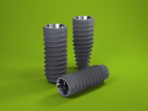
Tooth 21 was extracted carefully (Fig. 4), ensuring that the papillae and soft tissue contours remained intact. In order to minimize bone resorption and keep both the bone and soft tissue graft stable, a flap was not raised. Granulation tissue was removed and socket irrigation was performed with CHX2%. The implant bed was prepared for a ∅ 4.1 mm Straumann® BLT Implant (Roxolid® SLA® 4.1×12 mm). The implant was placed towards the palatal side of the socket, leaving a 2.5mm gap and 3.5mm in depth from our ideal gingival margin (Fig. 5). We prepared the provisional using a temporary long-term abutment and the patient’s own crown with the help of a silicone key to reposition the crown in its original exact location. The gap was filled with xenogenic bone graft, and we placed a connective tissue graft from the anterior palate with an envelope technique. The envelope was prepared with a sclerotome (sharp point). The temporary crown was screwed, and all occlusal contacts during centric and eccentric movements were eliminated (Fig. 6). Sutures were removed after 10 days (Figs. 7,8).
Prosthetic procedure
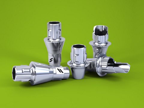
Osseointegration was checked after 10 weeks (Figs. 9,10). A customized impression transfer was created following the provisional contour (Figs. 11-13). The lab prepared the final restoration using a veneered zirconia crown (GC Initial) on the Straumann® Variobase® abutment (Figs. 14-16).
Final Result
In our practice we believe that immediate implants offer numerous advantages over conventional implants in terms of treatment duration, number of surgeries, morbidity and esthetic outcome. In this particular case, the patient benefited both from the standpoint of time and esthetic outcome (Figs. 17,18). We believe that it is not necessary to keep the buccal wall intact in order to place an immediate implant as, in most cases where the buccal wall is present, it will resorb within a short period of time as it is usually formed from bundle bone exclusively. We already know from the literature that bundle bone is dependent on the periodontal ligament and will disappear following extraction of the tooth. We believe that non-resorbed bone peaks from the neighboring teeth, followed by a good surgical technique with soft and hard tissue graft and immediate provisionalization, will guide the healing in a proper way to produce predictable results.


