Placement of a 2.9 mm Straumann® Bone Level Tapered implant in the area of a lateral maxillary incisor
A clinical case report by Mario Roccuzzo, Italy
This case describes a prosthetic restoration in the area of the lateral maxillary incisor by means of the Straumann® Bone Level/Bone Level Tapered implants with SLActive® surface. Furthermore, bone and connecting tissue grafting procedures were applied to ensure an appropriate implant performance and to achieve an excellent esthetic result.
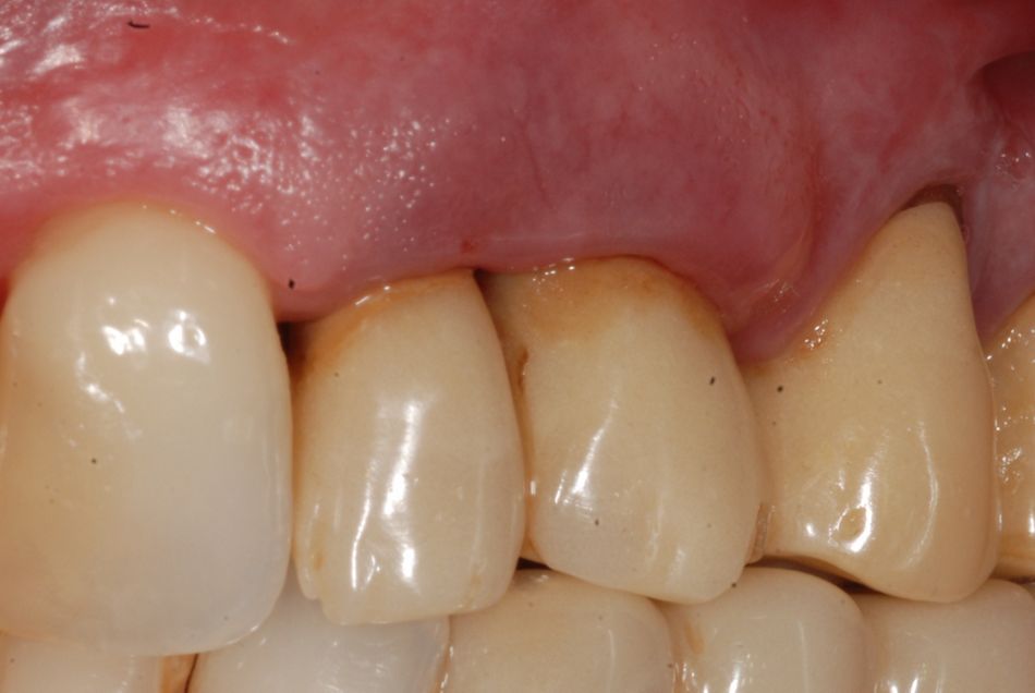
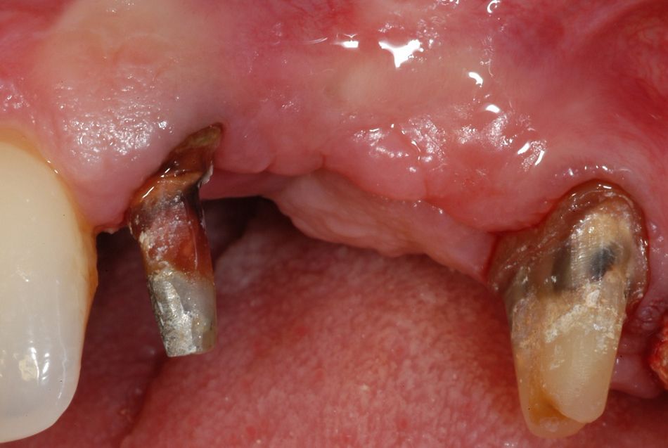
In July 2012, a 52-year-old, non-smoking female patient came for a consultation regarding her periodontal status of her left maxillary area (Fig. 1). She reported pain and frequent abscesses and expressed the desire to improve her esthetics and function. The patient presented several periodontal problems, including multiple gingival recessions and a non-plaque-related mucosal disorder. A thorough analysis was carried out and, following the initial periodontal treatment, the patient gave her informed consent for the extraction of 2.2 and 2.3, which were considered not worth treating.
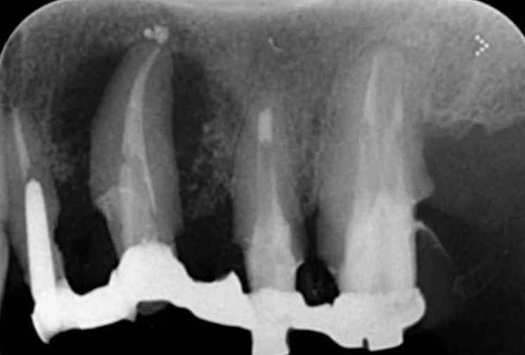
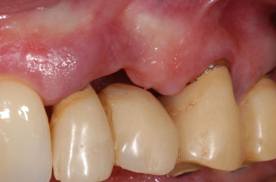
In order to minimize the discomfort during treatment, a step-by-step treatment was requested. The first task was to extract the canine, which was surrounded by a large radiolucent area (Fig. 2). Twelve weeks later, a substantial reduction in the horizontal and vertical dimensions of the crest was evident. The upper left lateral incisor was temporarily left in place to facilitate the positioning of a fixed provisional restoration (Fig. 3).
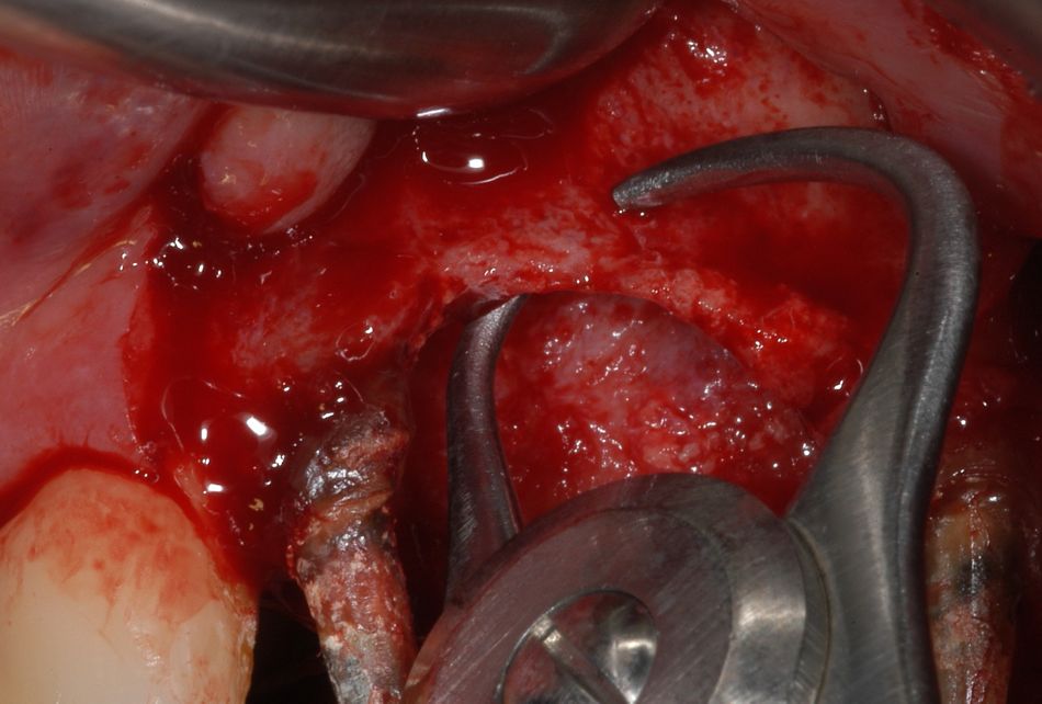
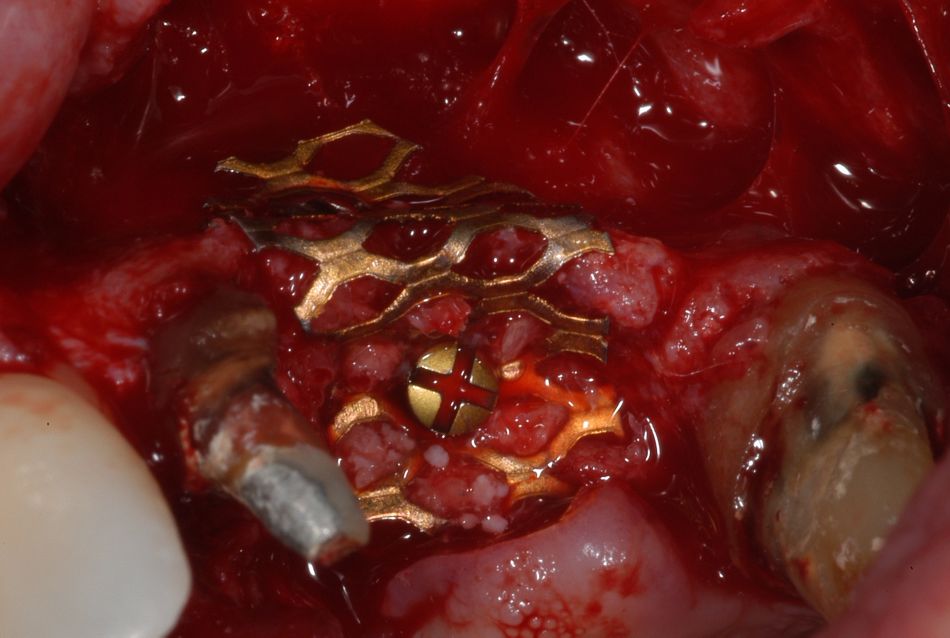
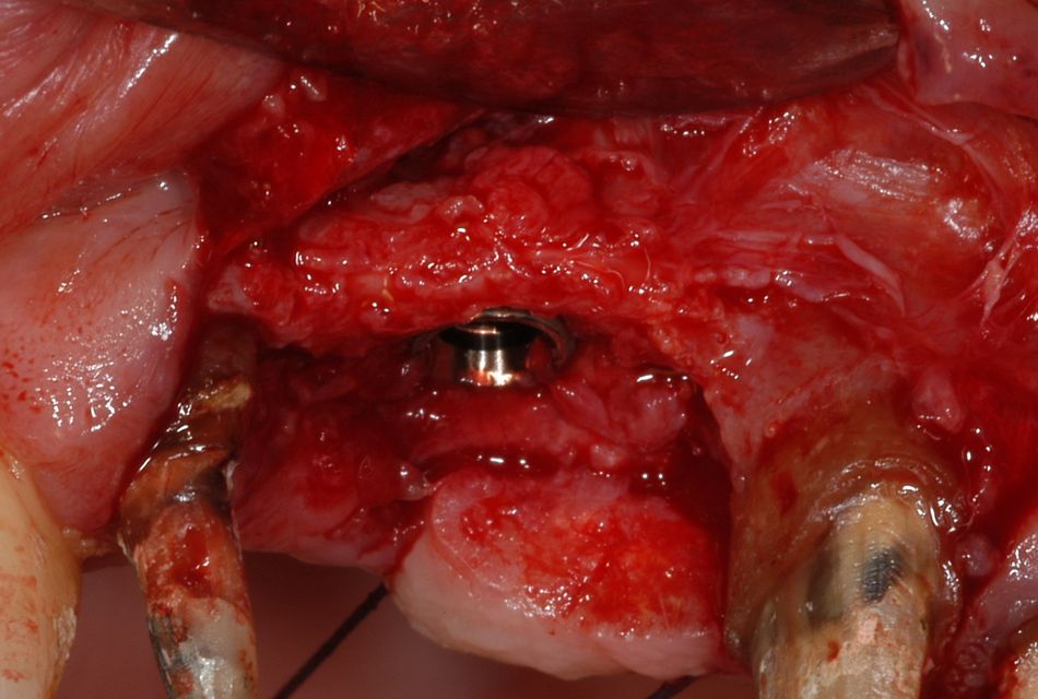
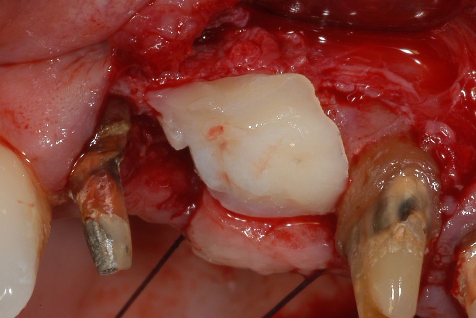
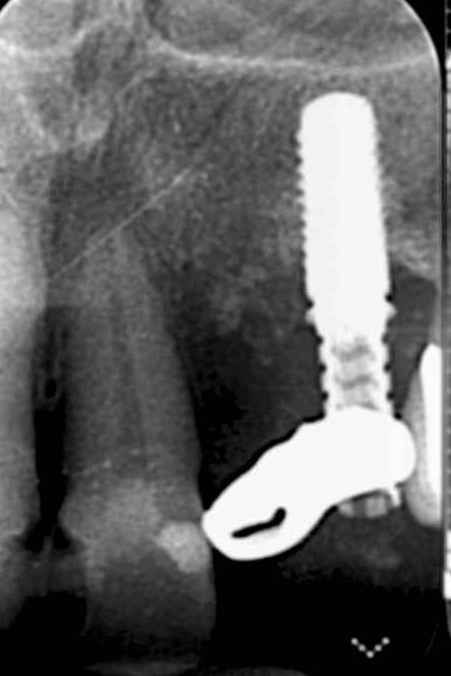
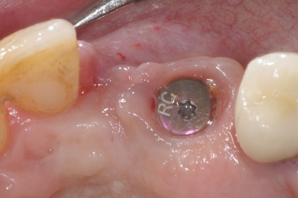
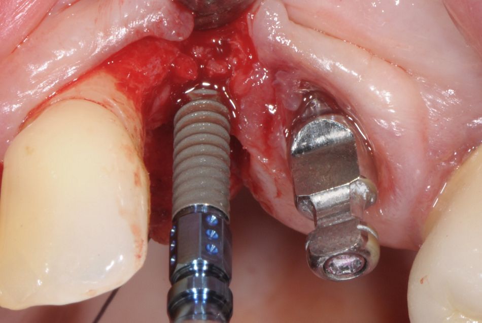
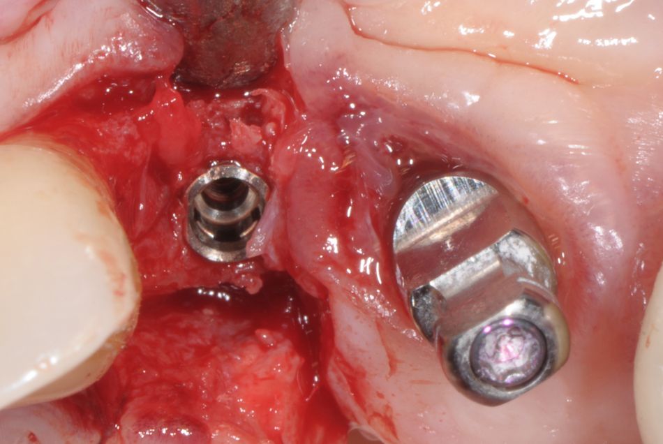
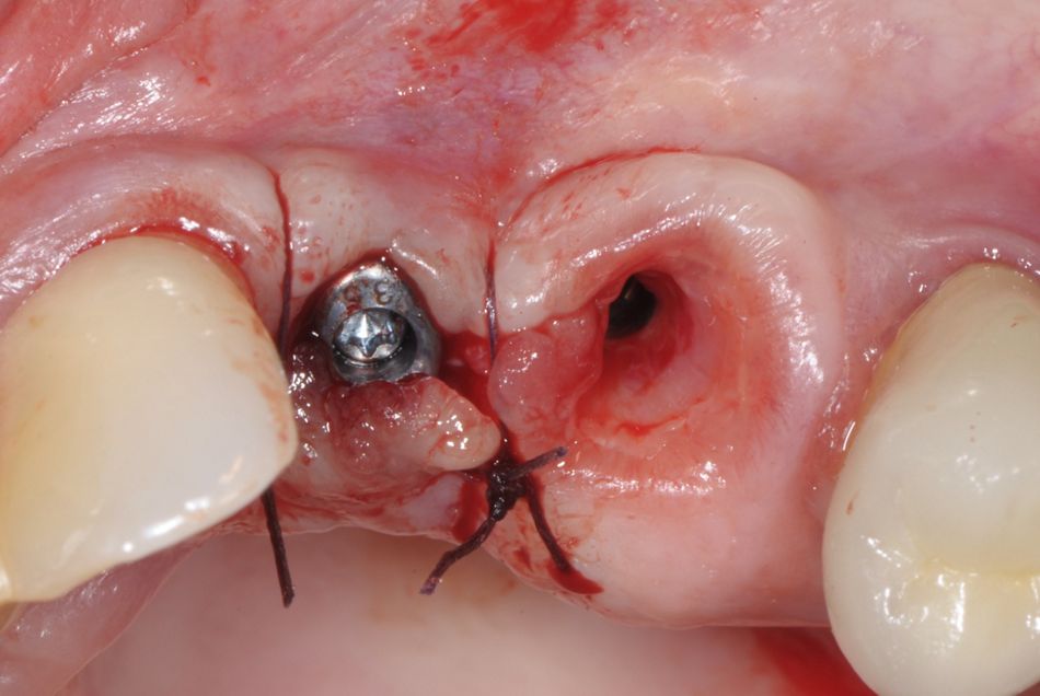
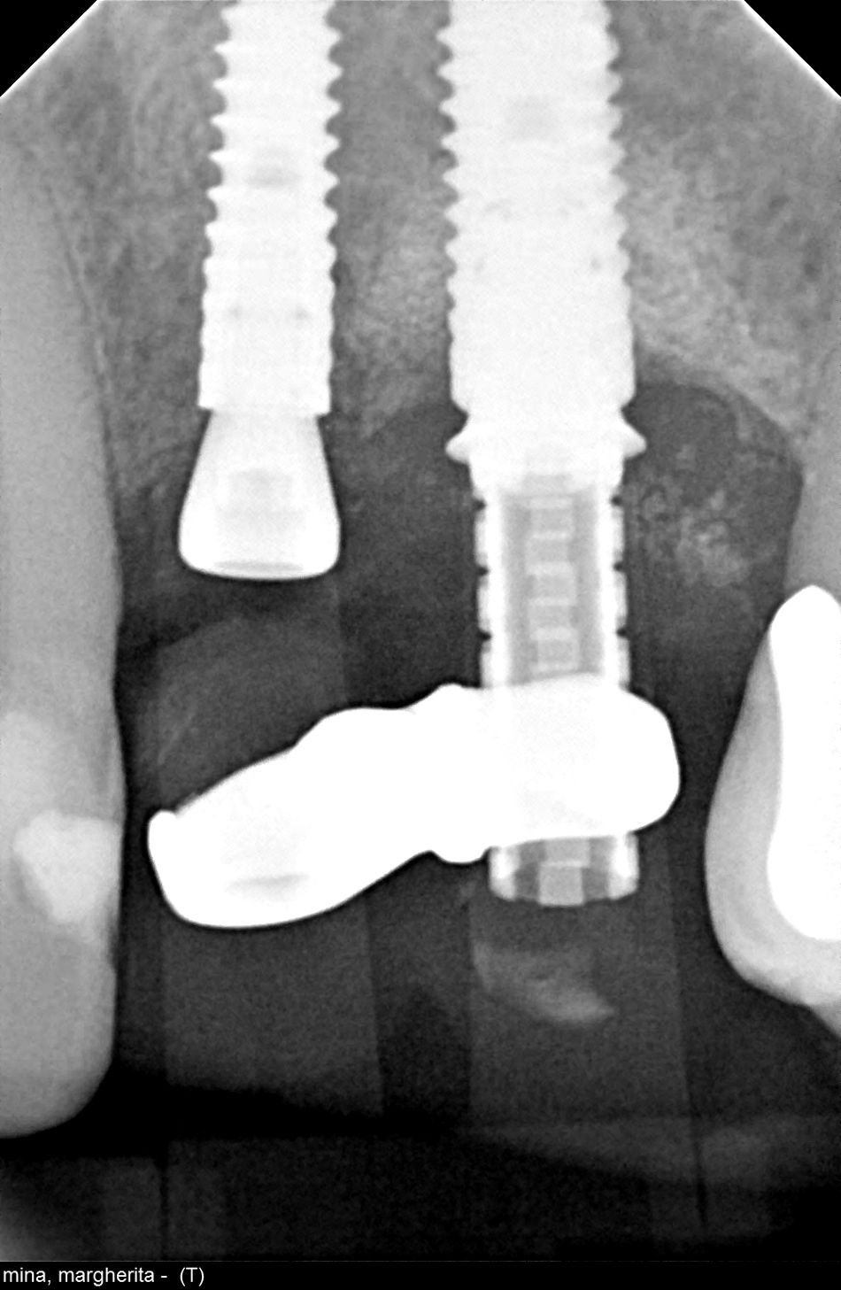
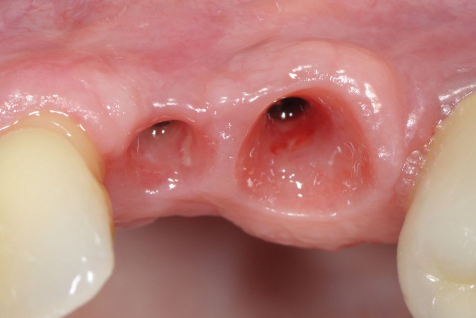
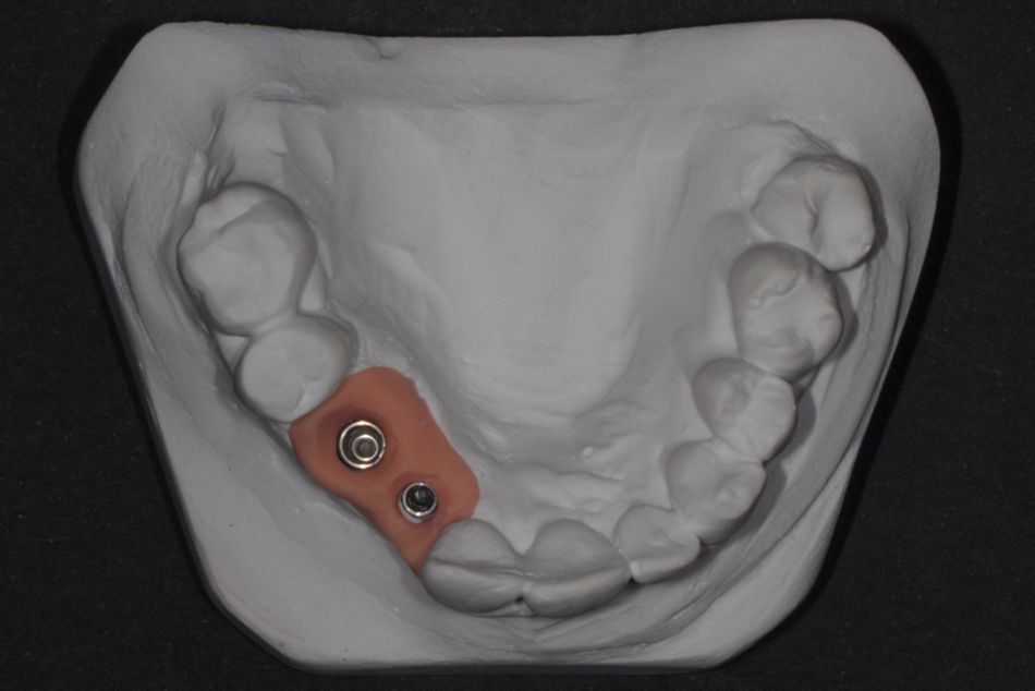
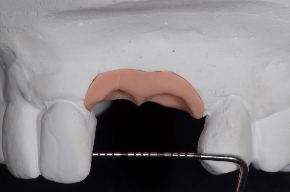
In January 2013, a full-thickness flap was raised to expose the bone. The severe atrophy did not allow implant placement and required a bone augmentation procedure (Fig. 4). Bone was harvested from the ramus and angle of the mandible using a device specifically designed for this purpose (Micross, Meta Reggio Emilia, Italy). A 0.2 mm thick Ti-Mesh was used to stabilize and protect the graft, as described previously by Roccuzzo et al. (2007, 2016) (Fig. 5). In September 2013, after 9 months of undisturbed healing, the flap was raised and measurement revealed a new width of the crest that was adequate for a regular implant diameter. Therefore, a BL, Ø 4.1 mm, SLActive® 10 mm, was used (Fig. 7). A connective tissue graft was harvested from the maxillary tuberosity and applied on top to increase the soft tissue volume (Fig. 8). A provisional fixed restoration with a mesial cantilever was adapted after the extraction of 2.2 (Figs. 9, 10). In May 2016, a Bone Level Tapered (BLT), Ø 2.9 mm, SLActive® 12 mm, Roxolid® implant was placed according to the manufacturer’s instructions. The implant was inserted in a self-tapping fashion, and primary stability was achieved (Figs. 11, 12). The use of a reduced-diameter implant resulted in more bone around the implant for better long-term success. The implant was placed at the center of the alveolar bone crest, leaving sufficient space both in relation to the tooth and the adjacent implant. The suturing technique employed ensured that the implant was not submerged (Fig. 13). The x-ray confirmed the proper positioning of the implant (Fig. 14). Three months after surgery, the peri-implant mucosa appeared free of inflammation (Fig. 15). The patient was then instructed in the correct brushing technique for ensuring adequate plaque control without injuring the soft tissues. After soft tissue maturation, an impression was taken and the cast prepared (Figs. 16, 17).
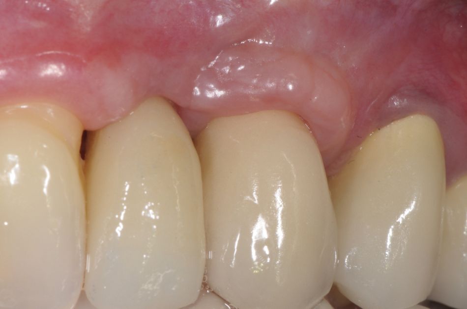
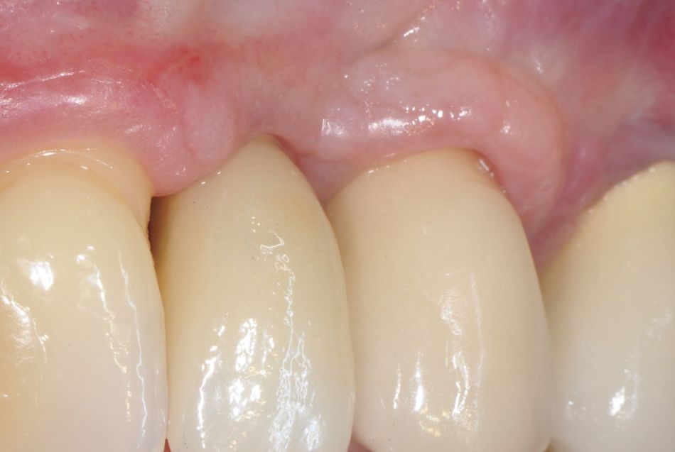
The two screw-retained crowns were delivered in October 2016 (Fig. 18), and the patient was asked to follow an individualized supportive periodontal therapy (SPT) program for an appropriate clinical and radiographic follow-up.
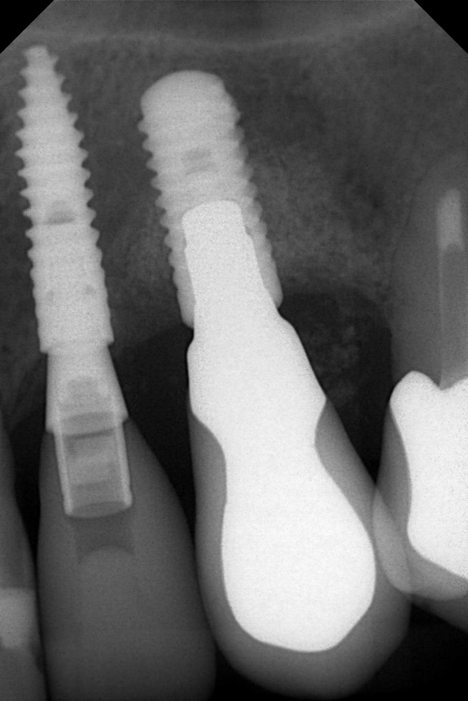
In April 2017, the radiographic examination confirmed the correct fitting of the crowns on the implants and the good level of the interproximal bone crest (Fig. 19). Plaque control was adequate, pocket depth was physiological and there was no bleeding on probing (Fig. 20).