Digital full-arch implant rehabilitation using the Straumann Pro Arch™ concept and Straumann BLC™ implants in a patient following head and neck radiotherapy
Introduction
Dental implant rehabilitation in patients who have undergone head and neck radiotherapy presents one of the most complex challenges in oral rehabilitation (1). Radiation therapy causes permanent alterations in the microvascular and cellular environment of bone and soft tissue, resulting in hypoxia, hypovascularity, and hypocellularity (2). These changes impair normal bone turnover and diminish the capacity for tissue repair, leading to delayed wound healing and an increased risk of infection, poor osseointegration, and osteoradionecrosis (ORN) (3). The degree of these complications is directly related to the total radiation dose, fractionation schedule, and time elapsed since treatment, as well as patient-related factors such as systemic health and nutritional status.
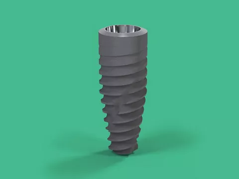
In irradiated bone, the compromised vascular network limits the recruitment of osteogenic and endothelial cells essential for osseointegration. This necessitates the use of implant systems that promote rapid and stable bone integration despite reduced biological potential. The Straumann BLC™ implant system offers biomechanical and surface characteristics that address these clinical demands. Its Roxolid® titanium-zirconium alloy provides enhanced tensile and fatigue strength compared with conventional pure titanium, permitting the use of narrower implants and thus minimizing surgical trauma to already compromised bone (4). Furthermore, the SLActive® surface possesses a nanostructured, hydrophilic topography designed to accelerate early bone healing (5). Experimental evidence demonstrates that the SLActive® surface enhances the adsorption of plasma proteins and promotes faster activation of platelets and osteogenic cells, resulting in increased early bone-to-implant contact. These effects are particularly advantageous in irradiated tissue, where the healing response is delayed and vascular perfusion is reduced. From a biomechanical standpoint, the tapered macro-geometry and slim apical design of the BLC implant enable accurate placement with high primary stability while minimizing compression of fragile cortical bone. The internal TorcFit™ connection also seals the implant–abutment interface, reducing micro gap-related inflammation and marginal bone loss. For patients who have undergone head and neck radiotherapy, these properties together can create a biologically and mechanically favorable environment for osseointegration. When applied through a computer-guided, minimally invasive surgical protocol, the Straumann BLC™ system can enhance surgical accuracy, preserve residual vascularized bone, and reduce intraoperative trauma.
The present clinical case report describes the use of the Straumann Pro Arch™ concept and Straumann BLC™ implants in the full-arch rehabilitation of a patient with a history of head and neck carcinoma treated with surgery and radiotherapy. The case illustrates how careful implant selection and digital planning can mitigate radiation-related risks and achieve stable functional and esthetic outcomes in a medically compromised individual.
Initial situation
Patient Background
The patient, a 62-year-old male, presented seeking comprehensive rehabilitation of his severely deteriorated dentition. His medical history was notable for head and neck squamous cell carcinoma of undetermined primary origin, for which he had undergone a right-sided selective neck dissection and adjuvant external beam radiation therapy approximately four years earlier. He was also being followed for a thoracic aortic aneurysm measuring 4.2 centimeters, managed with medical therapy and regular imaging surveillance. Additional conditions included gastroesophageal reflux disease and a cervical spinal fusion. The patient was systemically stable and reported no tobacco or alcohol use. His chief complaint was the inability to chew solid food. He had been on a soft diet for several years due to extensive dental breakdown and was seeking a fixed solution that would restore his ability to eat comfortably and improve esthetics.
Clinical Findings
Extraoral examination revealed no signs of swelling, erythema, or asymmetry. The patient exhibited a normal mandibular range of motion, with no trismus or joint tenderness (Figs. 1a-d). Intraoral examination and panoramic radiograph showed multiple crowns with recurrent decay, several non-restorable root canal–treated teeth, and generalized cervical caries (Figs. 2a,b, Fig. 1e). The gingival and mucosal tissues appeared healthy, and both the maxillary and mandibular ridges exhibited adequate volume and keratinized gingiva suitable for implant placement.
A panoramic radiograph demonstrated bilaterally symmetrical temporomandibular joints, clear maxillary sinuses, and extensive cervical radiolucencies around the remaining teeth (Fig. 1e). The previously existing implant at site #12 was clinically and radiographically stable, with no evidence of inflammation. The diagnosis was terminal dentition with poor prognosis for the remaining teeth and functional impairment secondary to radiation-related changes and dental destruction.
Fig. 1. Extraoral photos.
Fig. 2. Digital teeth arrangement using Smilecloud


Treatment planning
Given the patient’s oncologic history and desire for fixed prostheses, a digitally guided, minimally invasive, full-arch implant rehabilitation was planned. The approach aimed to preserve bone vascularity, minimize surgical trauma, and utilize implant systems with good performance in compromised bone conditions.
Smile Design on Smilecloud
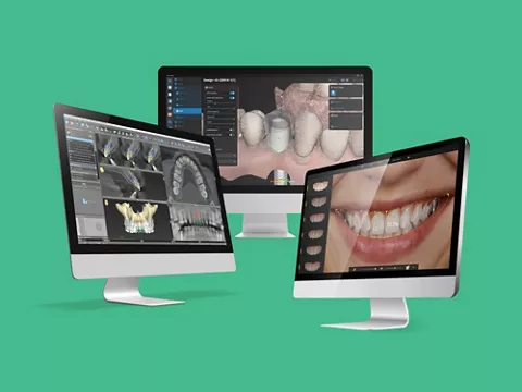
Digital planning began with a comprehensive esthetic and functional assessment using Smilecloud (Smilecloud Inc., Romania), a cloud-based interdisciplinary platform that integrates facial, dental, and radiologic data into a unified three-dimensional environment. First, the stack tool was used to synchronize multiple data sets including two-dimensional photographs, intraoral scans (IOSs), and cone beam computed tomography (CBCT) 3D radiographic data (Fig. 2c). Following this, the structure tool was selected to choose what teeth should be reshaped or removed (Fig. 2d). After that, an AI-assisted smile simulation was performed to determine optimal tooth proportions, incisal edge position, and occlusal plane orientation in harmony with the patient’s facial soft-tissue envelope (Fig. 2e). Teeth shapes and positions were modified on the 3D model and confirmed on the patient’s photo, allowing for real-time adjustments (Fig. 2f). The “YES” tool was also used to create an AI-generated video of the 3D smile design that will allow the patient to visualize the final outcome before starting the treatment (Video 2g). The resulting digital mock-up represented the intended prosthetic outcome and provided an esthetic reference for implant positioning. The completed smile design was exported in STL format to be imported into coDiagnostiX® (Version 10.8; Dental Wings Inc., Switzerland) for implant planning.
Implant planning on CoDiagnostix® (Figs. 3a-i and Figs. 4a-h)
CBCT data were first imported into coDiagnostiX® (Version 10.8; Dental Wings Inc., Switzerland). Digital IOSs of both arches were subsequently imported and registered to the CBCT dataset. The software’s artificial intelligence-assisted segmentation feature was activated to enable automated identification of anatomical structures (bone, teeth, and nerves) and automatic alignment of the IOS to the CBCT volume. This AI-driven fusion reduced manual error and ensured high-accuracy registration of the soft-tissue and hard-tissue datasets. The esthetic smile design previously generated in Smilecloud was then imported directly into coDiagnostiX®. Because Smilecloud exports files within a consistent 3D coordinate system, automatic alignment of the smile design to the CBCT-IOS dataset was achieved without the need for additional registration steps. This seamless interoperability preserved the spatial relationship between the prosthetic design and the anatomic structures, allowing prosthetically driven implant positioning.
For the maxillary arch, the pre-existing implant at site #12 was confirmed to be fully osseointegrated and was therefore retained. Three additional implants were planned in a four-implant full-arch configuration using Straumann BLC™ Roxolid® SLActive® implants (3.75 mm × 12 mm) at sites #15, #22, and #25. This configuration was selected to avoid sinus-floor elevation or bone-augmentation procedures, which could compromise healing in irradiated bone. Implant positioning was optimized to ensure full-arch support while eliminating the need for grafting, preserving both native bone and vascular integrity. Following virtual implant placement, screw-retained abutments (SRAs) were digitally incorporated to correct implant angulation and verify prosthetic accessibility where 17° angled SRAs were planned for sites #15 and #25, while a straight SRA was selected for site #22. Virtual verification confirmed that all screw-access channels emerged through the occlusal surfaces of the planned prosthesis. Selected teeth were then virtually extracted using the coDiagnostiX® automatic extraction tool. A mucosa- and tooth-supported guide was designed with anchor pins for additional stabilization. This design enabled flapless, minimally invasive implant placement, minimizing intraoperative trauma while enhancing biological and esthetic predictability. A similar digital workflow was applied to the mandible. Four Straumann BLC™ Roxolid SLActive implants (3.75 mm × 12 mm) were planned in a four-implant full-arch configuration at sites #35, #32, #42, and #45. The mandibular guide was designed to be mucosa-supported with anchor pins, ensuring stability and precision during flapless surgery.
For the interim prosthesis, the digital smile design exported from Smilecloud was re-utilized as the prosthetic reference. Extensions corresponding to the fixation-pin locations were incorporated to facilitate accurate intraoral pickup during surgery. All surgical guides and interim prostheses were exported in STL format. The surgical guides were fabricated using a 3D printer (Straumann® P30) and surgical guide resin (P Pro Surgical Guide 1000 g), while the temporary full-arch prosthesis was milled from polymethyl methacrylate (PMMA) using a 5-axis milling unit . Surgical and drilling protocols were automatically generated and exported in PDF format.
Fig. 3. Planning of maxillary arch implants and interim prosthesis on CoDiagnostix® software.
Fig. 4. Planning of mandibular arch implants and interim prosthesis on CoDiagnostix® Software.
Surgical procedure
Given the patient’s history of head and neck radiotherapy and the associated risk of radiation-induced fibrosis and ORN, a PENTO protocol (pentoxifylline and tocopherol) was initiated prior to the surgical phase. This pharmacologic regimen is supported by evidence demonstrating improved tissue oxygenation, reduction in radiation-induced fibrosis, and enhanced wound healing in irradiated tissues (6).
The patient was prescribed pentoxifylline 400 mg twice daily (BID) and vitamin E (tocopherol) 1000 IU daily, commencing four weeks before implant surgery and continuing for eight weeks postoperatively. Pentoxifylline, a methylxanthine derivative, acts as a hemorheologic and anti-TNF agent that increases erythrocyte flexibility and microvascular perfusion, thereby improving oxygen delivery to hypoxic irradiated tissues (6). Tocopherol functions as a potent antioxidant, scavenging reactive oxygen species and mitigating the chronic inflammatory and fibrotic responses that characterize late radiation injury. The synergistic action of these agents has been shown to partially reverse radiation-induced fibroatrophic changes in soft and hard tissues (6).
The patient received preoperative antibiotic prophylaxis consisting of amoxicillin 2 g orally one hour prior to surgery, along with a 0.12% chlorhexidine gluconate rinse for one minute to reduce intraoral microbial load. Local anesthesia was achieved using 4% articaine with 1:100,000 epinephrine (Septodont Septocaine®; Henry Schein). The procedures were performed under aseptic conditions and followed a digitally guided, minimally invasive protocol specifically designed to preserve vascularized soft tissues and minimize trauma to irradiated bone.
The surgical procedure commenced in the mandibular arch. All non-restorable teeth and residual root fragments were carefully extracted in a minimally traumatic manner with minimal flap reflection to maintain periosteal perfusion. The mucosa-borne, pin-supported surgical guide, designed from the digital treatment plan, was seated and stabilized using three fixation pins. Sequential osteotomies were performed using the Straumann® VeloDrill™ system. Four Straumann BLC™ Roxolid® SLActive® implants (3.75 mm × 12 mm) were placed at sites corresponding to teeth #35, #32, #45, and #42. All implants achieved high primary stability, with insertion torque values exceeding 35 N·cm and an implant stability quotient (ISQ) above 70. Following implant insertion, preplanned SRAs were tightened and torqued to 35 N·cm. The surgical field was thoroughly irrigated, and mucosal tissues were approximated with resorbable sutures.
Attention was subsequently directed to the maxillary arch. To maintain guide stability, strategic extractions were performed in stages, preserving key reference teeth until after implant placement. The mucosa/tooth-borne guide was accurately positioned and fixed with anchor pins. Guided osteotomies were then completed using the Straumann® VeloDrill™ system under copious irrigation.
Three Straumann BLC™ Roxolid® SLActive® implants (3.75 mm × 12 mm) were placed at sites #15, #22, and #25, while the pre-existing implant at site #12 was confirmed to be fully osseointegrated and retained. After verifying high primary stability of all implants, SRAs were tightened and torqued to 35 N·cm. The remaining non-restorable teeth were then extracted, and soft tissues were sutured with resorbable sutures . No grafting procedure was performed for both arches to decrease the likelihood of delayed healing, infection, and compromised osseointegration, which are known risks in irradiated bone.
Prosthetic procedure
Immediate Loading Phase
Immediately following implant placement, the prosthetic pickup was performed chairside using the prefabricated milled interim full-arch prostheses (Fig. 5a). For both arches, temporary titanium cylinders were secured to the SRAs and isolated with a rubber dam to prevent contamination. The internal channels of the cylinders were sealed with medical polytetrafluoroethylene (PTFE) tape. The prefabricated provisional prosthesis was seated using fixation pins aligned with the guide’s anchor-pin positions to ensure a repeatable fit. Using dual-cured injectable resin, the prosthesis was picked up intraorally to capture the exact implant positions. After polymerization, the prostheses were removed, the intaglio surface and margins were refined, and the prostheses were finished and polished in the laboratory. The prostheses were reinserted and torqued to 15 Ncm. Screw-access channels were sealed with PTFE tape and temporary resin. Occlusal adjustment was performed to eliminate any premature contacts and to establish even, bilateral centric occlusion with smooth excursive guidance. The patient was instructed to maintain a soft diet throughout the healing process, and postoperative medications including antibiotics, nonsteroidal anti-inflammatory drugs, and chlorhexidine rinses were prescribed. Follow-up evaluations were performed every two weeks during the first three months to monitor soft tissue healing. Occlusion was also rechecked at each appointment to ensure the absence of excessive load transfer to individual implants. Minor occlusal adjustments were performed as needed to maintain a stable, even contact pattern throughout the healing period (Fig. 5b). Panoramic radiograph taken to verify seating of interim prosthesis (Fig. 5c).
Fig. 5. Prosthesis pickup.
Definitive Prosthetic Phase
At the three-month follow-up, the interim prostheses were removed to evaluate osseointegration and prepare for the definitive restorative phase. All implants were clinically stable, with no evidence of mobility, tenderness, or peri-implant soft-tissue inflammation. ISQ values were recorded, and all implants demonstrated ISQ readings above 70, confirming adequate secondary stability and successful osseointegration (Figs. 6a,b). Following verification, the temporary prostheses were tightened again, and pre-preparation scans of both the maxillary and mandibular arches were obtained using an intraoral scanner (3Shape Trios 3). These scans served as a digital reference for the laboratory technician to reproduce the established vertical dimension, occlusal scheme, and esthetic contours when designing the definitive restorations. The interim prostheses were then removed again to capture emergence profile scans of both arches, accurately documenting the soft-tissue contour developed during the healing phase. Metal scan bodies for SRAs were subsequently hand-tightened, and an additional scan was performed to register the three-dimensional implant positions. The interim prostheses were reinserted thereafter, and a digital bite registration was recorded (Figs. 7a-g). All digital records (implant positions, pre-preparation, emergence profiles, and occlusal registrations) were imported into the computer-aided design (CAD) software (exocad GmbH). The implant positions were identified by aligning the scanned scan bodies with their corresponding digital library components. Titanium bases for SRA abutments (Coping for Variobase® for Bridge/Bar Cylindrical for Screw-retained Abutments; Straumann) were selected as the titanium bases. The definitive maxillary and mandibular prostheses were designed based on the pre-preparation scans, ensuring the preservation of the functional and esthetic parameters established with the temporaries.
Fig. 6. Healing phase after 3 months of osseointegration.
Fig. 7 Digital implant impressions.
Parallel 3D-printed master models were fabricated, on which verification jigs were assembled to confirm passive fit and the accuracy of implant positioning prior to final milling (Figs. 8a-i). The designed frameworks were also 3D printed as prototypes for intraoral try-in. Titanium abutments were provisionally attached to the printed prototypes to verify the fit, occlusion, and esthetics intraorally. Once passivity and esthetic parameters were confirmed clinically and, on the models, the prototypes were copy-milled to fabricate the definitive restorations (Figs. 9a-e). Both arches were produced as full-arch monolithic zirconia prostheses with titanium abutments for SRA foundations . The definitive prostheses were CAD/CAM milled in monolithic zirconia , with the gingival portions characterized and stained to replicate natural mucosal esthetics. The Variobase® components were cemented to the intaglio surfaces following a standardized adhesive protocol: air abrasion with 50 µm Al₂O₃ particles, application of metal and zirconia primer followed by dual-cured cement (Figs. 10a-g). Upon delivery, the prostheses were seated intraorally and torqued to 15Ncm. (Figs. 10h,ih:) A panoramic radiograph confirmed complete seating and passive adaptation of both restorations (Fig. 10j). Occlusion was carefully refined to eliminate premature contacts and to ensure evenly distributed bilateral centric loading with smooth excursive movements.
Fig. 8. Verification of implant impressions.
Fig. 9. Prototype try in.
Fig. 10. Long-term prosthesis.


Treatment outcomes
At the two-week follow-up after delivery, the patient reported a marked improvement in oral function, regaining full masticatory efficiency and noting enhanced speech clarity and overall comfort. From an esthetic perspective, the final outcome met his expectations, exhibiting a natural smile line, balanced tooth proportions, and harmonious gingival contours that integrated seamlessly with his facial features (Fig. 10k). The patient expressed satisfaction with the rehabilitation, particularly the ability to chew and smile confidently after years of functional and social limitation. The patient was given a night guard to protect the definitive restorations and mitigate the effects of nocturnal parafunctional activity, thereby supporting the long-term stability and longevity of the rehabilitation (Fig. 10l).