#Preventive 13. Jun 2025

Reconstructive management of peri-implantitis defect using GalvoSurge® in a periodontally compromised patient

The case highlights the benefit of electrolytic decontamination with GalvoSurge®, along with guided bone regeneration (GBR) and supportive treatment, in maintaining the stability of soft and hard tissues around an implant affected by peri-implantitis.

Introduction

Peri-implantitis remains one of the most challenging biological complications in implant dentistry, particularly in patients with a history of periodontitis. Effective decontamination of the implant surface is critical to the successful management of this condition. Traditional mechanical and chemical methods often fall short in completely eliminating the biofilm from the complex microstructure of implant surfaces, especially in advanced cases.

GalvoSurge®, an electrolytic cleaning system, offers a novel and minimally invasive approach by generating hydrogen bubbles that physically lift biofilm and bacterial contaminants from the implant surface without damaging the titanium. This case report describes the successful management of peri-implantitis in a high-risk patient with a documented history of severe generalized periodontitis. Remarkably, the peri-implantitis developed 16 years after implant placement, emphasizing the need for long-term monitoring and effective treatment strategies in vulnerable patients.

The case highlights the pivotal role of electrolytic decontamination with GalvoSurge®, combined with guided bone regeneration (GBR) and personalized supportive care, in achieving soft and hard tissue stability around an implant affected by advanced peri-implantitis.

Initial situation

A 46-year-old male non-smoking patient with a diagnosis of severe generalized periodontitis was referred to our practice in January 2002 for an integral dental treatment.  The patient was initially professionally treated by scaling and root planing in order to obtain infection control. In addition, personalized oral hygiene instructions were given to ensure proper domiciliary plaque control. After the assurance of good motivation and compliance (FMPS < 20%; FMBS < 20%), the patient was prepared for the surgical phase.

The surgical phase included guided tissue regeneration with amelogenin (Straumann® Emdogain®) in locations where deep periodontal infrabony defects were present.

Nine months after the surgical phase, an orthodontic treatment, aiming at correcting malposition, creating contact points, and giving a non-traumatic occlusion, was initiated.

After completion of the active orthodontic therapy, the patient was placed on an individually tailored maintenance care program, including continuous evaluation of the occurrence and the risk of disease progression. In addition, motivation, reinstruction, instrumentation, and treatment of re-infected sites were performed as needed.

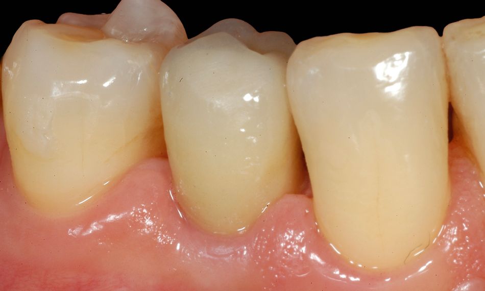
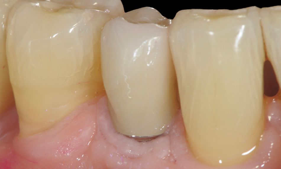
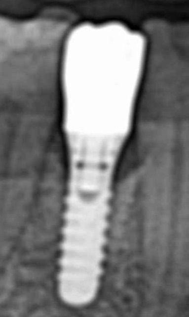
In November 2006 a tissue-level implant (SP, Ø 4.1 mm RN, 12 mm) (Institut Straumann AG, Basel Switzerland) was placed to replace a hopeless premolar in site #44 (Fig. 1). After a three-month healing period, the patient was recalled for the prosthetic phase and a ceramic crown was cemented (Fig. 2).

The patient was seen regularly for supportive maintenance care, 2-3 times a year as needed.

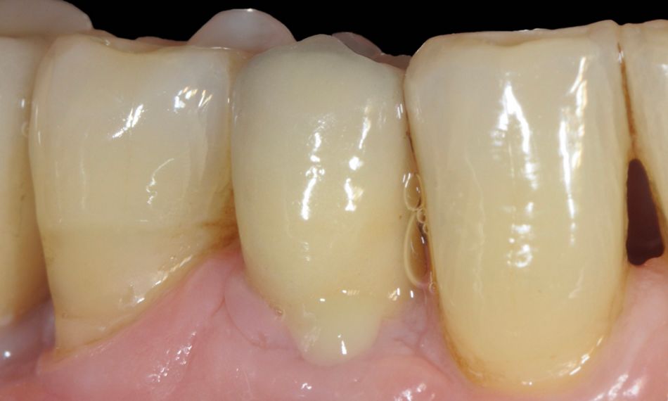
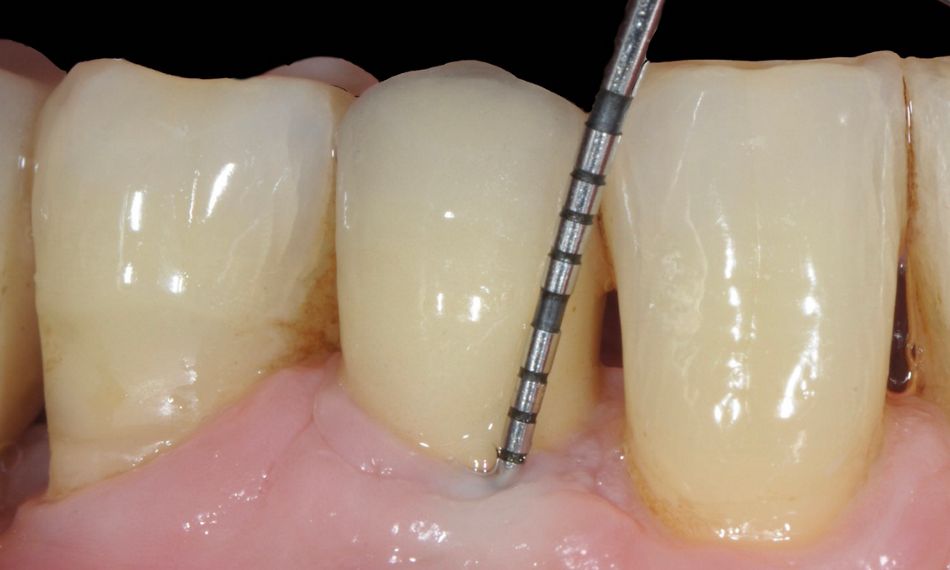
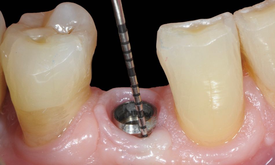
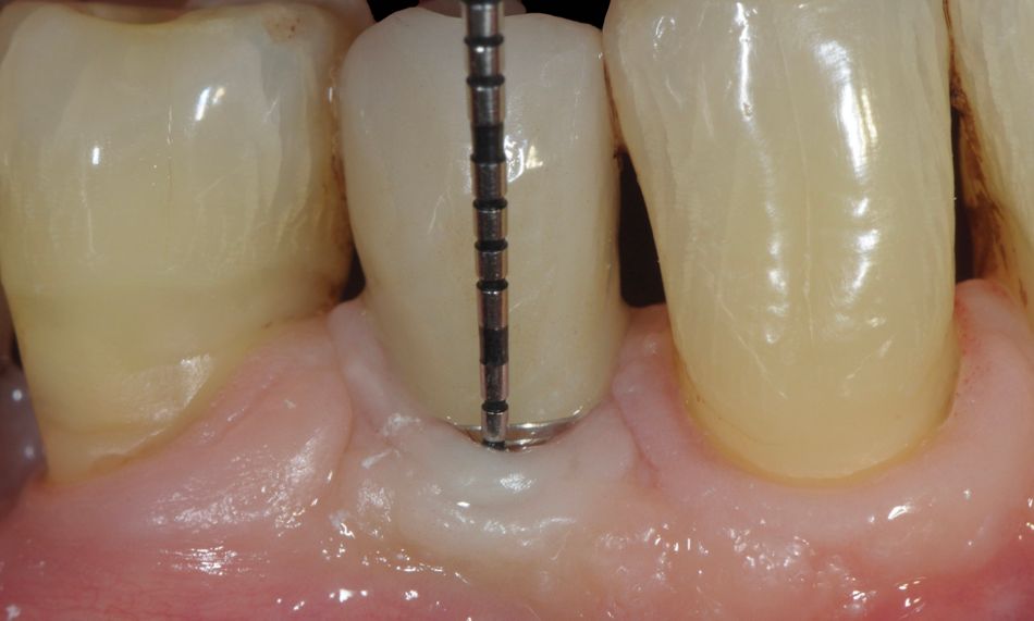
In February 2022, he came to the office complaining of discomfort and tenderness in the lower right region, which had been going on for several weeks. This seemed to be related to the implant in position #44 (placed 16 years before), where a deep pocket and abundant suppuration on probing were observed (Figs. 3, 4).

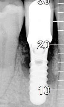
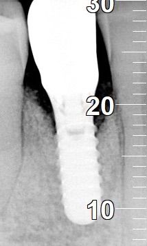
The radiograph confirmed the presence of a deep crater-like bone defect around the implant in #44, and a diagnosis of peri-implantitis was confirmed (Fig. 5).

Treatment planning

The treatment workflow included:

  1. Non-surgical periodontal therapy.
  2. Reconstructive surgical treatment with GalvoSurge®.
  3. Final prosthetic rehabilitation with a new screw-retained crown.
  4. Follow-up appointments for monitoring.

Surgical procedure

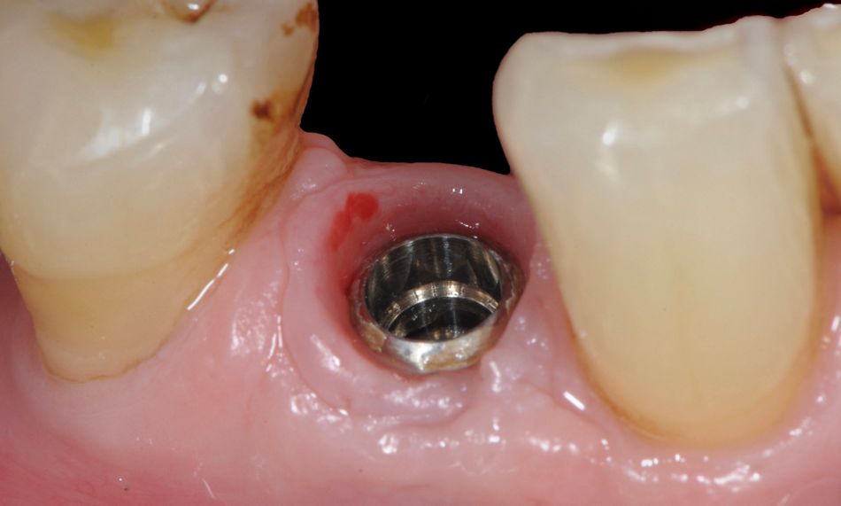
In order to facilitate access, the ceramic crown and the abutment were removed. The clinical picture demonstrated a minimal accumulation of plaque deposits in the region. Moreover, the adjacent teeth were free from signs of periodontitis (Fig. 6).

The area selected for surgery was anesthetized with mepivacaine plus epinephrine 1:100,000.

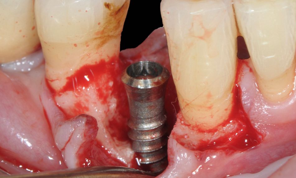
A full-thickness flap was elevated buccally and lingually around the implant. Due to the significant probing depth, a minimal oblique vertical incision was added mesially to the canine to have full access to the button of the defect. Subsequently, all granulation tissue was completely removed from the defect area, and the implant surfaces were thoroughly debrided using titanium brushes (Fig. 7).

GalvoSurge® (Institut Straumann AG, Basel, Switzerland) was utilized to decontaminate the surface of the implant. GalvoSurge® is an electrolytic cleaning device that works by applying a mild electric current through an electrolytic solution, generating hydrogen bubbles that lift and remove biofilm and bacteria from the implant surface without damaging it (Fig. 8).

In light of the available evidence, a dual approach combining mechanical and chemical decontamination was done before evaluating the configuration of the peri-implant bone defect. The surface was finally rinsed thoroughly with sterile saline solution.

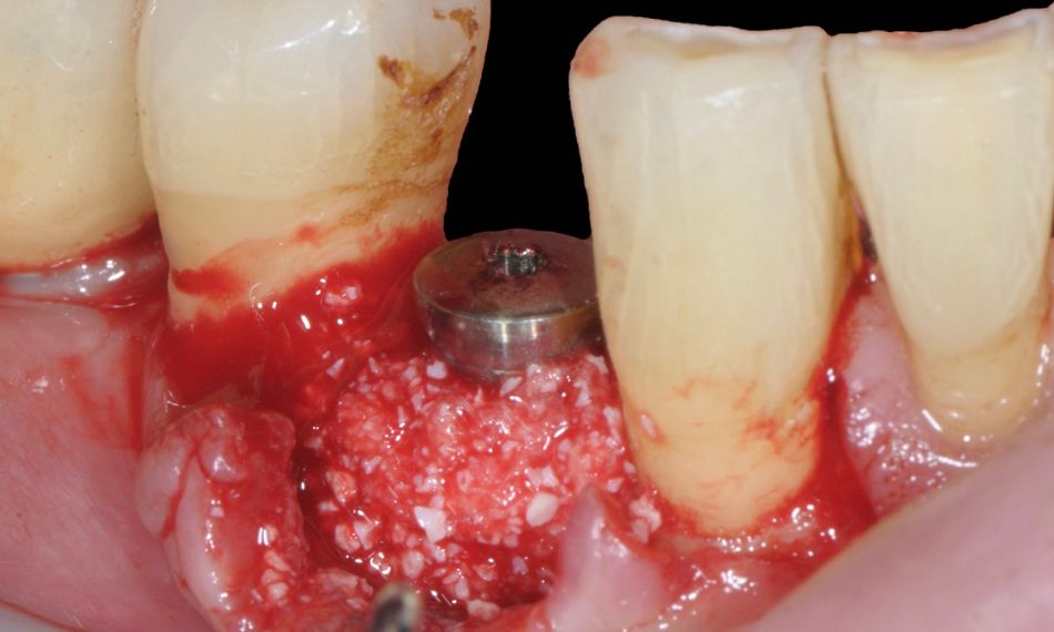
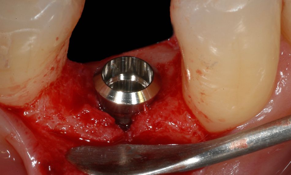
The defect was filled with deproteinized bovine bone mineral with 10% collagen. Care was taken not to overfill the defect and to stay slightly underneath the ideal crestal margin to facilitate placement of a Connective Tissue Graft (CTG) (Fig. 9).

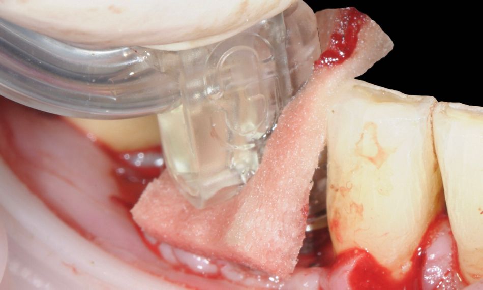
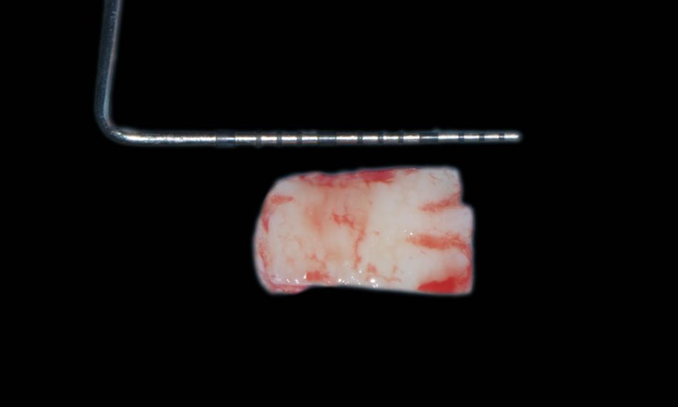
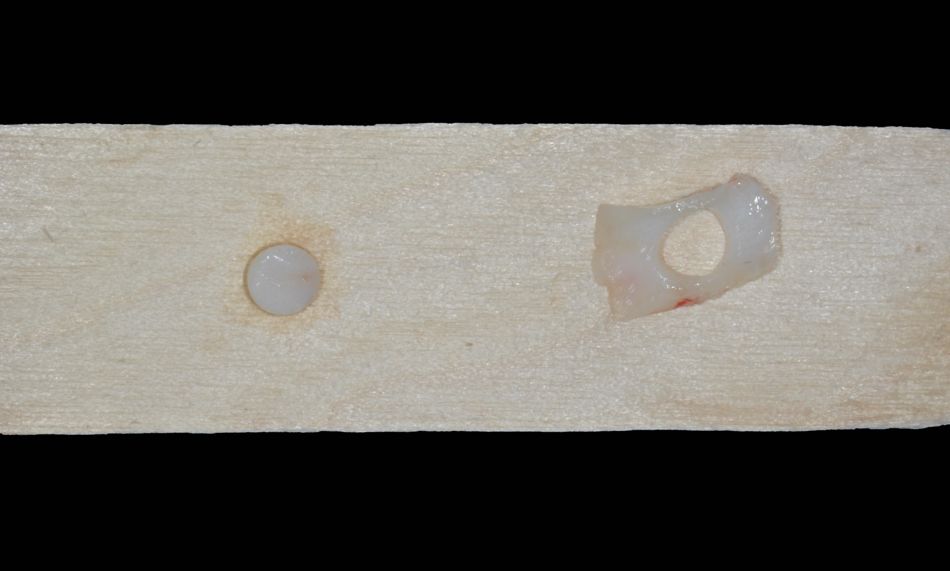
A thick tissue graft was excised from the palate, de-epithelialized with a blade, and trimmed with a mucotome to ensure optimal adaptation to the collar of the implant, which measured 4.1 mm in diameter. The thickness of the mucosal graft was approximately 2 mm. A punch was performed on the tissue to allow it to adapt better in the implant area (Figs. 10,11).

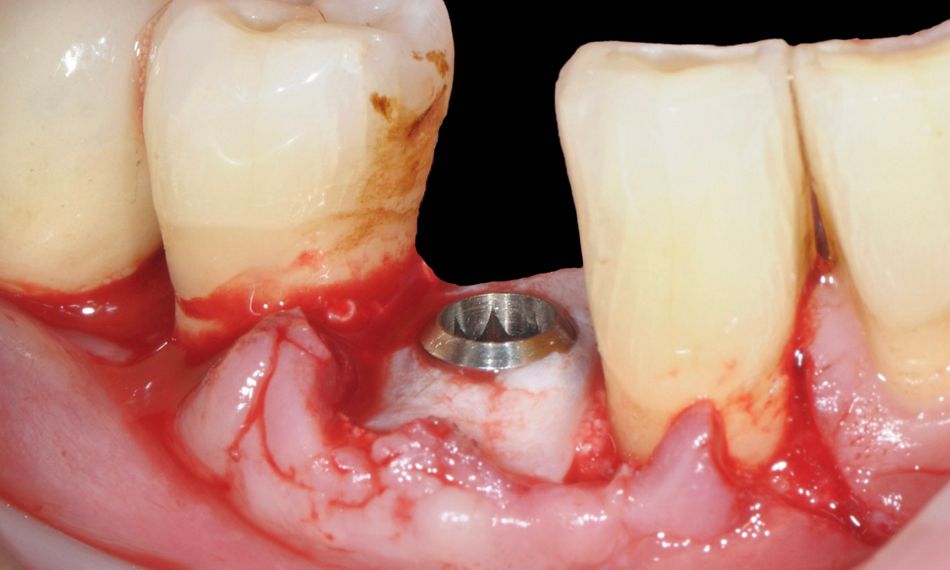
The prepared connective tissue graft was adapted so as to create a collar around the implant and ensure stability of the grafted material (Fig. 12).

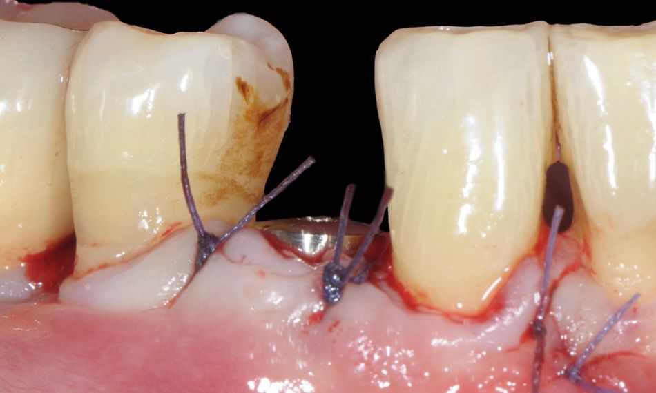
Finally, the flap was repositioned coronally and fixed with sutures to ensure an optimal non-submerged healing (Fig. 13).

Immediately after surgery, the patient was advised to apply an ice pack over the treated area and continue this for at least four hours. He was instructed to avoid tooth brushing and trauma to the surgical site for three weeks and to rinse with 0.12% chlorhexidine digluconate for one minute three times a day for the same period.

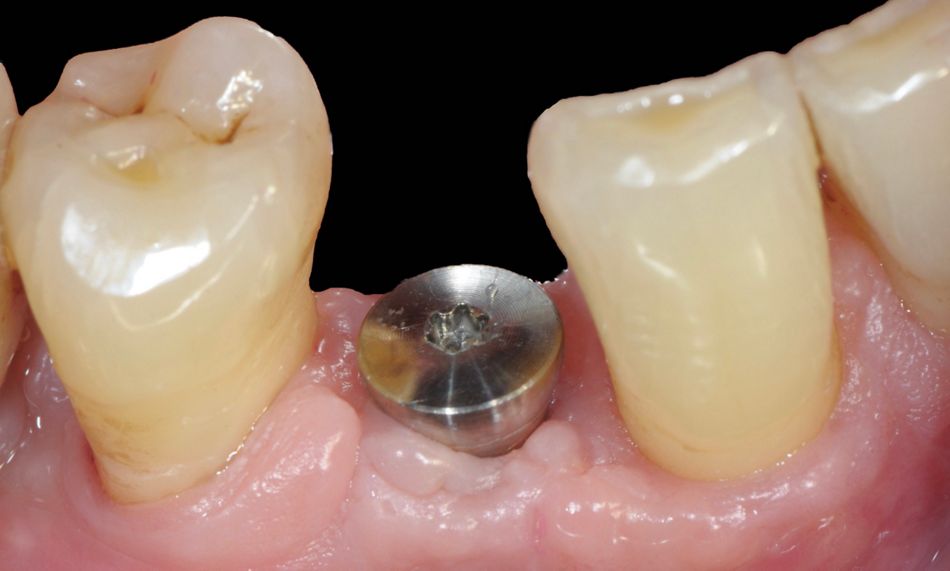
The patient was seen after 7 days to monitor healing and after 14 days for suture removal. Subsequently, weekly follow-ups were done for the first 4 weeks. At the 4-week follow-up, healing had progressed without complications (Fig. 14).

Afterward, the patient was seen every 3 months during the first year, and every 6 months in subsequent years.

Prosthetic procedure

Motivation, reinforcement of oral hygiene, and instrumentation were performed as needed. The 1-year post-op clinical picture revealed complete soft tissue coverage of the rough surface of the implant, the absence of inflammation and bleeding on probing, and shallow pockets (Fig. 15). A new screw-retained ceramic crown was delivered (Fig. 16).
The intraoral radiograph revealed significant improvement of the interproximal bone levels (Fig. 17).

The last clinical and radiographic examination was carried out in November 2024, 22 years after the initial visit, and confirmed the stability of both the periodontal and the peri-implant tissues (Figs. 18,19).

Treatment outcomes

The use of GalvoSurge® during bone reconstructive surgery contributed to the successful re-establishment of peri-implant health.

A personalized Supportive Peri‐implant/Periodontal Care (SPC) program played a crucial role in maintaining these outcomes, ensuring continuous risk assessment and long-term success.

This approach highlights the effectiveness of GalvoSurge® in combination with a reconstructive surgical approach.

Author’s testimonial

With this patient’s history in mind, it is advisable to place implants with a long-term vision so that maintenance is easy both for the patient and the dentist. Therefore, in order to reduce the risk of biological complications, patients should be placed on a personalized SPC program that includes continuous evaluation of the risk of disease progression. On the other hand, if peri-implantitis is detected around properly placed implants, it can be successfully treated by means of the described technique in a high percentage of cases, even in severe cases such as the one presented here.