Surfaces du Straumann® Dental Implant System 

Redéfinir les limites de ce qui est possible.

Acheter maintenant

De SLA®, la surface de référence dans le domaine, à la surface implantaire SLActive® haute performance, Straumann a toujours placé la barre plus haut pour établir de nouveaux standard dans l’ostéointégration. ZLA®, développé pour l’implant Straumann® PURE Ceramic, perpétue cette tradition.

Images SEM sur le réseau de fibrine, image fournie avec l’aimable autorisation d’Empa, 2016

Straumann® SLActive®

Performance au-delà de l’imagination.

Acheter maintenant

SLActive® est la surface haute performance de Straumann présentant un potentiel de cicatrisation important. Elle est caractérisée par une forte prédictibilité et une ostéointégration accélérée1-8. Des études récentes démontrent une excellente performance clinique des implants SLActive® même dans les protocoles de traitement les plus difficiles et chez des patients présentant des problèmes de santé.9-11

Cherchez-vous à obtenir plus d’informations ? Vous les trouverez dans le Centre de téléchargement.

Prédictibilité

Taux de survie des implants en mise en charge immédiate après 10 ans : 98,2 %.9

Ostéointégration

Temps de cicatrisation réduit passant de 6 à 8 semaines à 3 à 4 semaines dans toutes les indications.*

Biologie

Meilleure génération osseuse même au niveau de sites compromis.12

Performance

Taux de survie des implants après 5 ans chez les patients irradiés présentant des lésions osseuses : 100 %.10,11

* par rapport à SLA®. Indications : d’un espace unitaire à une mâchoire édentée.

Images SEM sur le réseau de fibrine, image fournie avec l’aimable autorisation d’Empa, 2016

Straumann® SLA®

Longévité et efficacité dans la pratique quotidienne.

Acheter maintenant

La surface classique SLA®, introduite en 1998, est basée sur une technique de sablage à gros grains qui génère une macro-rugosité sur la surface en titane. Ceci est suivi d’une attaque à l’acide qui superpose une micro-rugosité. La topographie résultante offre une structure idéale pour l’attachement cellulaire. De nombreuses études cliniques et précliniques évaluées par des pairs ont confirmé ses fortes performances à long terme et sa fiabilité, ce qui en fait l’une des surfaces les mieux documentées en implantologie dentaire. Selon une étude indépendante, l’odds ratio de développer une péri-implantite était plus de trois fois plus élevé chez les patients traités par des systèmes implantaires concurrents par rapport aux implants Straumann® avec la surface SLA®.13

Cherchez-vous à obtenir plus d’informations ? Vous les trouverez dans le Centre de téléchargement.

Taux de survie

Taux de survie élevés et fiables entre 95,1 % et 98,8 % documentés par différentes études après 5 et 10 ans de suivi.14-20

Préservation osseuse

Perte osseuse moyenne de 0,5-1 mm après 10 ans (valeurs initiales définies comme étant le moment de mise en charge de l’implant).15,16

Faible prévalence de péri-implantite

Très faible prévalence de la péri-implantite (1,8 %) sur une période de 10 ans.15
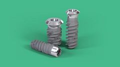
Straumann® ZLA®

Une surface avec des caractéristiques d’ostéointégration révolutionnaires.

Acheter maintenant

Straumann® ZLA® est la surface du système implantaire Straumann® PURE Ceramic et présente des caractéristiques d’ostéointégration révolutionnaires qui sont équivalentes à la surface Straumann® SLA®.

Structure

Similaire à SLA® en termes de macro et micro rugosité.

Ostéointégration

Temps de cicatrisation comparable à celui de SLA®21-23

Esthétique

Fixation moins importante de la plaque – un facteur important pour la réussite à long terme de l’implant23.

Histoires présentées sur youTooth

Aucun résultat pour votre recherche

Erreur pendant le téléchargement des articles

Explorer plus d’articles youTooth

Téléchargements

Documents

Vidéos et Animations

Preuves scientifiques

Catalogues

Bibliographie

SLActive® 1 Straumann SLActive implants compared to Straumann SLA implants. Lang NP, Salvi GE, Huynh-Ba G, Ivanovski S, Donos N, Bosshardt DD. Early osseointegration to hydrophilic and hydrophobic implant surfaces in humans. Clin Oral Implants Res. 2011 Apr;22(4):349-56. doi: 10.1111/j.1600-0501.2011.02172.x. 2 Rupp F, Scheideler L, Olshanska N, de Wild M, Wieland M, Geis-Gerstorfer J. Enhancing surface free energy and hydrophilicity through chemical modification of microstructured titanium implant surfaces. Journal of Biomedical Materials Research A, 76(2):323-334, 2006. 3 De Wild M. Superhydrophilic SLActive® implants. Straumann document 151.52, 2005; Katharina Maniura. Laboratory for Materials – Biology Interactions Empa, St. Gallen, Switzerland, Protein and blood adsorption on Ti and TiZr implants as a model for osseointegration. EAO 22nd Annual Scientific Meeting, October 17 – 19 2013, Dublin. Kopf BS, Schipanski A, Rottmar M, Berner S, Maniura-Weber K, Enhanced differentiation of human osteoblasts on Ti surfaces pre-treated with human whole blood. Acta Biomaterialia. 2015 June; 19: 180-190. Kopf BS, Ruch S, Berner S, Spencer ND, Maniura-Weber K, The role of nanostructures and hydrophilicity in osseointegration: In-vitro protein-adsorption and blood-interaction studies. J Biomed Mater Res A. 2015 August; 103 (8): 2661-2672. 4 Schwarz, F., et al., Bone regeneration in dehiscence-type defects at non-submerged and submerged chemically modified (SLActive®) and conventional SLA® titanium implants: an immunohistochemical study in dogs. J Clin. Periodontol. 35.1 (2008): 64-75. 5 Rausch-fan X, Qu Z, Wieland M, Matejka M, Schedle A. Differentiation and cytokine synthesis of human alveolar osteoblasts compared to osteoblast-like cells (MG63) in response to titanium surfaces. Dental Materials 2008 Jan;24(1):102-10. Epub 2007 Apr 27. 6 Schwarz F, Herten M, Sager M, Wieland M, Dard M, Becker J. Histological and immunohistochemical analysis of initial and early osseous integration at chemically modified and conventional SLA® titanium implants: Preliminary results of a pilot study in dogs. Clinical Oral Implants Research, 11(4): 481-488, 2007. 7 Raghavendra S, Wood MC, Taylor TD. Int. J. Oral Maxillofac. Implants. 2005 May–Jun;20(3):425–31. 9. 8 Oates TW, Valderrama P, Bischof M, Nedir R, Jones A, Simpson J, Toutenburg H, Cochran DL. Enhanced implant stability with a chemically modified SLA® surface: a randomized pilot study. Int. J. Oral Maxillofac. Implants. 2007;22(5):755–760. 9 Nicolau P, Guerra F, Reis R, Krafft T, Benz K , Jackowski J 10-year results from a randomized controlled multicenter study with immediately and early loaded SLActive implants in posterior jaws. Presented at 25th Annual Scientific Meeting of the European Association of Osseointegration – 29 Sep – 1 Oct 2016, Paris. 10 Nelson, K., Stricker, A., Raguse, J.-D. and Nahles, S. (2016), Rehabilitation of irradiated patients with chemically modified and conventional SLA implants: a clinical clarification. J Oral Rehabil, 43: 871–872. doi:10.1111/joor.12434. 11 Patients treated with dental implants after surgery and radio-chemotherapy of oral cancer. Heberer S, Kilic S, Hossamo J, Raguse J-D, Nelson K. Rehabilitation of irradiated patients with modified and conventional sandblasted, acid-etched implants: preliminary results of a split-mouth study. Clin. Oral Impl. Res. 22, 2011; 546–551. 12 Straumann (2016). SLActive® supports enhanced bone formation in a minipig surgical GBR model with coronal circumferential defects. Unpublished data. SLA® 13 Buser D, Janner SF, Wittneben JG, Bragger U, Ramseier CA, Salvi GE. 10-year survival and success rates of 511 titanium implants with a sandblasted and acid-etched surface: a retrospective study in 303 partially edentulous patients. Clin Implant Dent Relat Res. 2012 Dec;14(6):839-51. 14 Fischer K, Stenberg T.: Prospective 10-year cohort study based on a randomized controlled trial (RCT) on implant-supported full-arch maxillary prostheses. Part 1: sandblasted and acid-etched implants and mucosal tissue. Clin Implant Dent Relat Res. 2012 Dec;14(6):808-15. 15 van Velzen FJ, Ofec R, Schulten EA, Ten Bruggenkate CM,.10-year survival rate and the incidence of peri-implant disease of 374 titanium dental implants with a SLA surface: a prospective cohort study in 177 fully and partially edentulous patients. Clin Oral Implants Res. 2015 Oct;26(10):1121-8. 16 Cochran DL, Jackson JM, Bernard JP, ten Bruggenkate CM, Buser D, Taylor TD, Weingart D, Schoolfield JD, Jones AA, Oates TW Jr. A 5-year prospective multicenter study of early loaded titanium implants with a sandblasted and acid-etched surface. Int J Oral Maxillofac Implants. 2011 Nov-Dec;26(6):1324-32. 17 Cochran D, Oates T, Morton D, Jones A, Buser D, Peters F. Clinical field trial examining an implant with a sand-blasted, acid-etched surface. J Periodontol. 2007 Jun;78(6):974-82. 18 Bornstein MM, Schmid B, Belser UC, Lussi A, Buser D. Early loading of non-submerged titanium implants with a sandblasted and acid-etched surface. 5-year results of a prospective study in partially edentulous patients. Clin Oral Implants Res. 2005 Dec;16(6):631-8. 19 Roccuzzo M1, Aglietta M, Bunino M, Bonino L. Early loading of sandblasted and acid-etched implants: a randomized-controlled double-blind split-mouth study. Five-year results. Clin Oral Implants Res. 2008 Feb;19(2):148-52. 20 Derks J, Schaller D, Håkansson J, Wennström JL, Tomasi C, Berglundh T. Effectiveness of Implant Therapy Analyzed in a Swedish Population: Prevalence of Peri-implantitis. J Dent Res. 2016 Jan;95(1):43-9. doi ZLA® 21 Bormann KH, Gellrich NC, Kniha H, Dard M, Wieland M, Gahlert M. Biomechanical evaluation of a microstructured zirconia implant by a removal torque comparison with a standard Ti-SLA implant. Clin Oral Implants Res. 2012 Oct;23(10):1210-6. doi: 10.1111/j.1600-0501.2011.02291.x. Epub 2011 Nov 14. 22 Gahlert M, Roehling S, Sprecher CM, Kniha H, Milz S, Bormann K. In vivo performance of zirconia and titanium implants: a histomorphometric study in mini pig maxillae. Clin Oral Implants Res. 2012 Mar;23(3):281-6. doi: 10.1111/j.1600-0501.2011.02157.x. Epub 2011 Aug 2. 23 Gahlert M1, Röhling S, Wieland M, Eichhorn S, Küchenhoff H, Kniha H A comparison study of the osseointegration of zirconia and titanium dental implants. A biomechanical evaluation in the maxilla of pigs. Clin Implant Dent Relat Res. 2010 Dec;12(4):297-305. doi: 10.1111/j.1708-8208.2009.00168.x.

ZLA® 23 Roehling S, Astasov-Frauenhoffer M, Hauser-Gerspach, Braissant O, Woelfler H, et a., In Vitro Biofilm Formation On Titanium And Zirconia Implant Surfaces, J Periodontol. 2016 Oct 7:1-16. [Epub ahead of print] DOI: 10.1902/jop.2016.160245