Meet increasing patient expectations
With the increasing patient expectations and awareness, it is vital to keep offering differentiated experiences. Engage patients by offering access to a unique digital restorative experience and tailored treatments solutions meeting all expectations as well as individual situations.
Nowadays, patients are more and more looking for comfortable treatment.
100% Of Patients prefer intraoral scanning because it's more comfortable and faster1.
61% From these patients think that it's more comfortable than conventional impression because it reduces:1
• Queasiness
• Breathing difficulty
• Teeth and Periodontal sensivity
• Discomfort during mouth was opened.
Dental practices offering intraoral scanner and CAD/CAM made restorations are growing and will continue to grow faster in the upcoming years2,3.
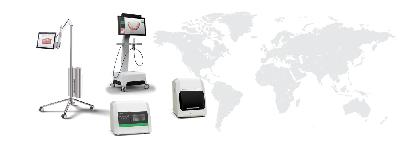
UNITED STATES

- 1 in 4 dental practicies perform intraoral scanning2
- 1 in 3,5 group practices use CAD/CAM milling systems2
EUROPE
- 1 in 8 dental practicies perform intraoral scanning
- 1 in 7 group practicies use CAD/CAM milling systems2
(1) YUZBASIOGLU, Emir et al. Comparison of digital and conventional impression techniques: evaluation of patients’ perception, treatment comfort, effectiveness and clinical outcomes. BMC oral health, v. 14, n. 1, p. 10, 2014.
(2) Digital Dental Solution Market Assessment – Voice of the customer – US and EU comparison summary, December 2017. For In house and milling center. Digital Dental Solution Market Assessment – Voice of the customer – US and EU comparison summary, December 2017
(3) CAD/CAM Dental Systems - Global Pipeline Analysis, Competitive Landscape and Market Forecast to 2017
Provide unique patient experience by a complete access to the digital restorative workflow
Neodent libraries are available for the main CAD/CAM softwares

CARES Visual

Exocad

Dental Wings

3shape
START PRECISELY
Treatment planning using intraoral scanner allows saving time and providing an optimal patient comfort. Neodent scan bodies have been designed to precisely secure the restorative position within a user-friendly experience.
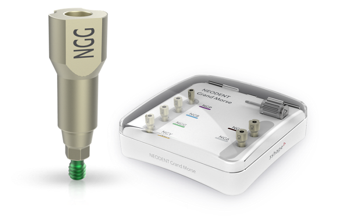
Neodent® IntraOral Scanbody
Neodent scan bodies are designed for an efficient and ease of digital impression use:
- Auto-recognition using CARES visual*
- Single patient use, sterile, peek-made
- For implant and abutment level impression
3Shape Scanbody for Neodent
The 3Shape scan bodies make your implant position transfer and system- identification simple:
- Auto-recognition using TRIOS*
- Multiple use
- Autoclavable titanium-made
- For implant level impression
FINISH WITH EFFICIENCY
3D printing dental model allow having predictable and repeatable results that provide ideal fit in less time. Additionally, it allow printing complex geometries with accurate detail visualization. Access to the highest level of realism with the Neodent hybrid analog.
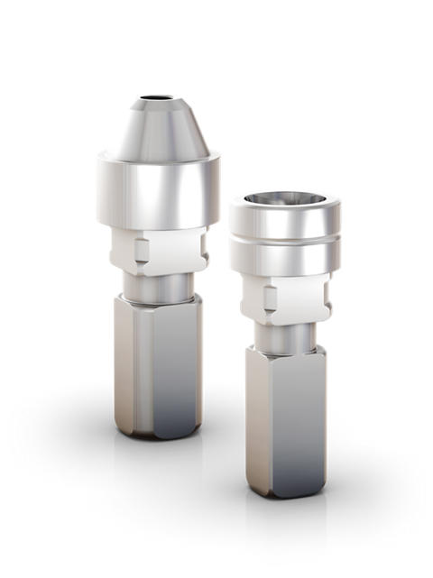
Hybrid Repositionable Analog
- For printed or plaster model
- Ant rotational with precise engaging positioning
- For implant and abutment level impression
How it connects?

1. DATA ACQUISITION
DIGITAL IMPRESSION
- Straumann CARES IntraOral Scanner
- 3Shape TRIOS
- Other IntraOral Scanner

2. PROSTHETIC DESIGN
CAD SOFTWARES
- CARES Visual
- 3Shape
- Dental Wings
- Exocad

3. MANUFACTURING
MILLING MACHINES
- CARES C Series
- CARES M Series
- ZirkonZahn M4
- Sirona Cerec MC XL
- Any other Milling Machine

4. FINISHING
3D PRINTERS
- CARES P Series 3D Printer
- Other 3D Printers
Meet patient expectations by offering a comprehensive restorative treatment options
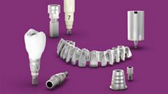
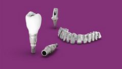
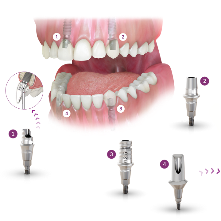
Mastering every step in every restorative workflow is essential for dental professionals. With Neodent deliver immediate natural-looking esthetic through a comprehensive restorative portfolio, covering all indications: from single to edentulous.
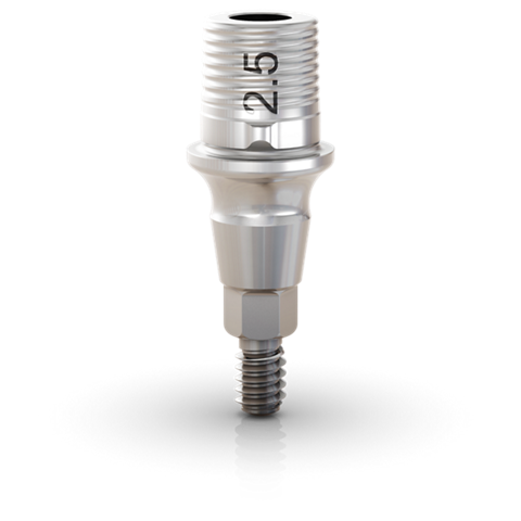
NEODENT TITANIUM BASES
- For single-unit restoration
- Several gingival heights (from 0.8 to 4.5 mm)
- Several abutment diameters (3.5, 4.5 and 5.5 and 6.5 mm)
- Allows crown customization and maximum angulation of 30°.
NEODENT TITANIUM BASES C
- For single-unit restoration
- For CEREC workflow
- Several gingival heights (from 0.8 to 4.5 mm)
- Allows crown customization and maximum angulation of 20°.
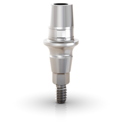
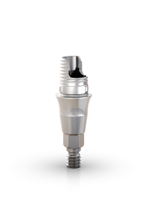
NEODENT TITANIUM BASES ANGLED SOLUTION
- For single-unit restoration with angled screw channel
- Several gingival heights (from 0.8 to 2.5 mm)
- Several abutment diameters (4.0, 4.5 and 5.5 mm)
- Allows maximum angulation of 25° depending on the gengival height and cementable area
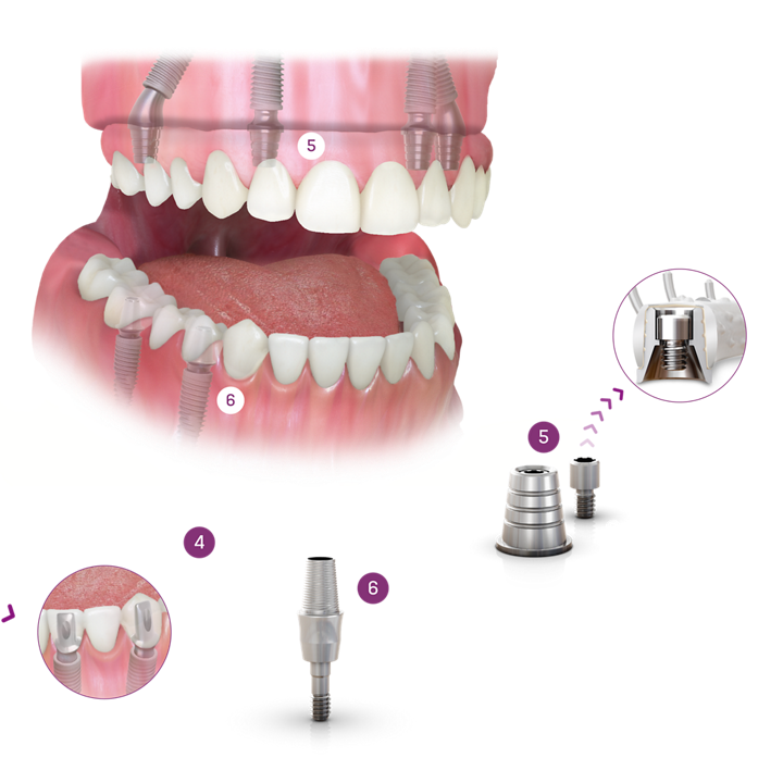
NEODENT TITANIUM BASES FOR BRIDGES
- For multi-unit restoration
- Several gingival heights (from 0.8 to 4.5 mm)
- Several abutment diameters (3.5, 4.5 and 5.5 mm)
- Allows maximum divergence of 10° for ø3.5 and 16° for ø4.5 and ø5.5
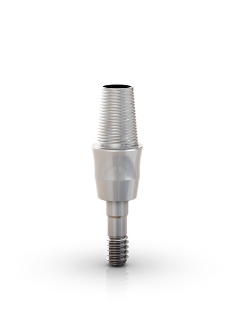
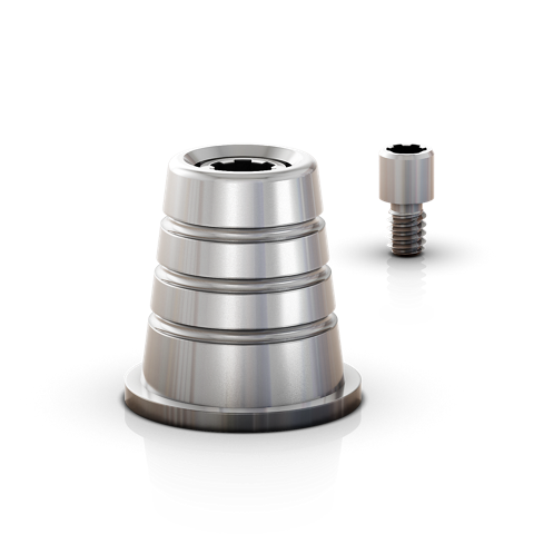
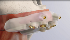
One Step Hybrid Coping:
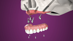
- For multi-unit or edentulous restorations
- For screw-retained bars and bridges
- Provides a passive fit
- Allows maximum divergence of 28°
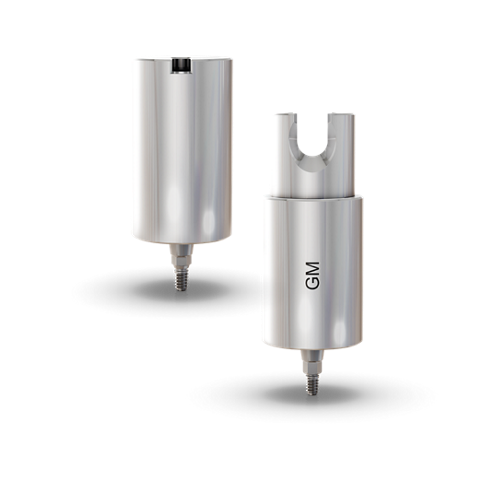
NEODENT TITANIUM BLOCK
- For single or multi-units restorations
- For Medentika holder in two different diameters: 11.5 and 15.8 mm
- For Amann Girrbach holder in one diameter: 12 mm
- Allows maximum angulation of 30°
