


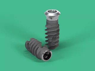
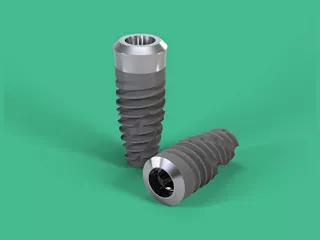
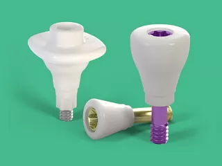
Straumann® Dental Implant System magas színvonalú megoldást kínál a fogpótlásra, különböző fogászati igények kielégítésére tervezett implantátumok széles választékát biztosítva. A pontos tervezéséről és fejlett anyagairól ismert rendszer titán és cirkónium-dioxid implantátumokat használ a biokompatibilitás, a tartósság és a kiváló csontintegráció érdekében.
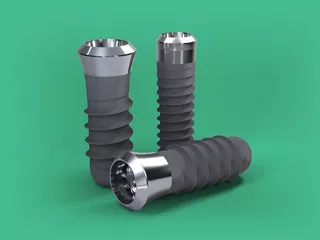
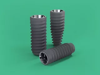
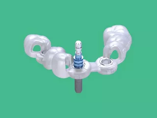
A Straumann implantátumai többféle méretben és konfigurációban állnak rendelkezésre, egyfogas, többfogas vagy teljes fogsoríves restaurációkhoz.
Az innovatív megoldások közé tartozik a fokozott tartósságot biztosító Roxolid® anyag1 és a SLActive® felületi technológia, amely felgyorsítja a gyógyulást és hosszú távú stabilitást biztosít.2


Kiemelt történetek a youToothon
Közelgő tanfolyamok
A rendszer áttekintése
További információra van szüksége? Itt megtalálja: Erőforrásközpont.