


Straumann® Regenerative Solutions is our contribution to regenerating hard and soft tissue around teeth and dental implants to maintain long-lasting function and esthetics. We are not just another supplier; we are here to support you by providing scientifically backed-up solutions for simple to complex cases around Tooth Preservation, Implant-Site Management and Implant Preservation.
Focus areas


Portfolio at a glance
Discover more
Clinical cases and scientific articles related to Straumann® Regenerative Solutions in our online magazine.


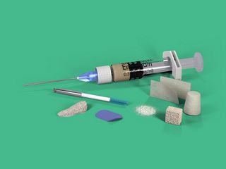
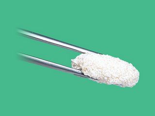
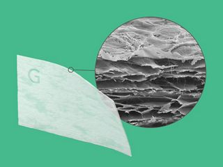
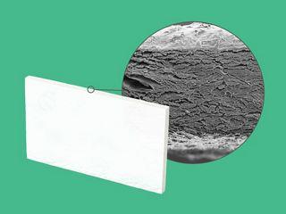
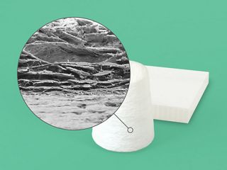
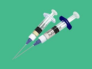
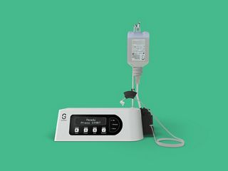
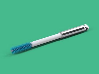
Straumann® Regenerative Solutions Product portfolio
Learn more about our Straumann® Regenerative Solutions portfolio.