


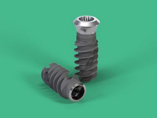
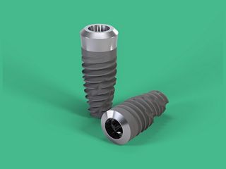
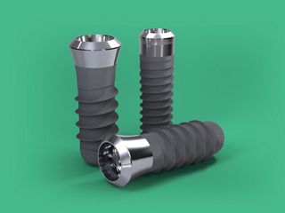
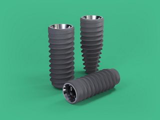
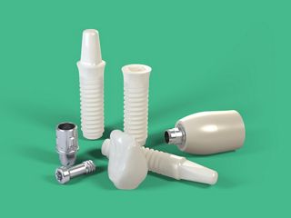
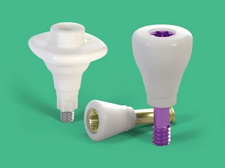
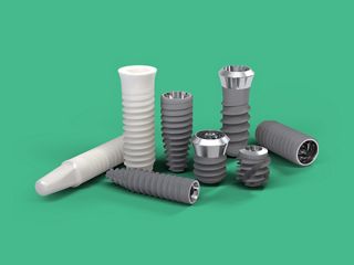
Le système implantaire dentaire Straumann® est une solution de haute qualité destinée au remplacement des dents et proposant une vaste gamme d’implants afin de répondre à diverses indications dentaires. Connu pour son ingénierie précise et ses matériaux avancés, le système utilise des implants en titane et en zircone assurant biocompatibilité, durabilité et une excellente ostéointégration.
Les implants Straumann sont disponibles en différentes tailles et configurations pour s’adapter aux cas de restaurations unitaires, plurales ou d’arcades complètes.
Leurs caractéristiques innovantes incluent le matériau Roxolid® pour sa résistance éprouvée1 ainsi que la surface SLActive®, qui accélère la cicatrisation et favorise une stabilité à long terme2.

Histoires présentées sur youTooth
Formations à venir
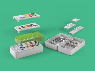
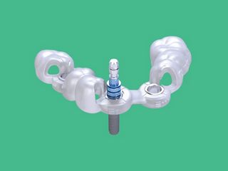
Présentation du système
Cherchez-vous à obtenir plus d’informations ? Vous les trouverez dans le Centre de ressource.