


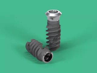
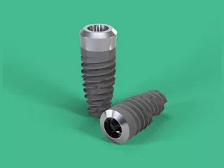
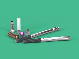
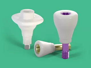
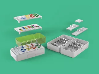
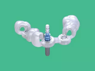
Straumann® Dental Implant System je vysoce kvalitní řešení pro zubní náhrady, které nabízí spektrum implantátů navržených pro různé stomatologické potřeby. Tento systém, známý svým přesným technickým zpracováním a pokročilými materiály, využívá titanové a zirkoniové implantáty pro biokompatibilitu, dlouhou životnost a vynikající osteointegraci.
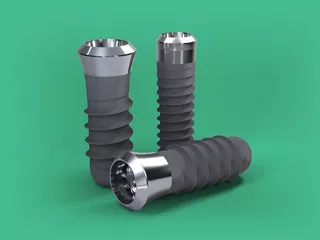
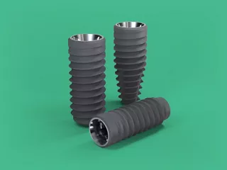
Implantáty Straumann jsou dostupné v různých velikostech a konfiguracích, pokrývajících náhrady jednoho zubu, více zubů i celkové náhrady.
Inovativní prvky zahrnují materiál Roxolid® pro zvýšenou pevnost1 a technologii povrchu SLActive®, která urychluje hojení a zajišťuje dlouhodobou stabilitu2.

Příběhy na youTooth
Chystané kurzy
Přehled systému
Hledáte další informace? Podívejte se na centrum materiálů.