





 International – English



Multidisciplinary Rehabilitation of a Maxillary Lateral Incisor in Regenerated, Low Density Bone, Using the Axiom® X3S Implant

Dr. Georgios Emmanouilidis
United Kingdom
Implantologist focused on oral surgery and guided surgery
- Graduated in Greece (2016) with postgraduate training in oral surgery, implant dentistry and sedation
- Clinical focus on guided implant surgery and efficient treatment protocols
- Anthogyr-UK Ambassador, involved in clinical education and case sharing
Introduction.
Implant placement in the aesthetic zone presents significant clinical challenges, particularly in the presence of unfavourable spacing and deficient ridge conditions. Achieving predictable primary stability while preserving peri-implant soft tissue architecture is especially demanding in sites requiring bone regeneration and multidisciplinary intervention.
In such contexts, treatment success relies on a prosthetic-driven approach, combining surgical precision, soft tissue management and optimized implant design to ensure both functional and aesthetic outcomes. Managing regenerated low-density bone (D3–D4) remains a key difficulty, requiring adapted protocols to achieve stability and long-term predictability.
This case illustrates how a multidisciplinary and digitally guided workflow, using the Axiom® X3S implant system, can address these challenges and support predictable rehabilitation in a complex anterior indication.
Case Presentation.
A 52-year-old female patient, medically fit and a non-smoker, presented with a non-restorable fractured upper right lateral incisor (UR2) with retained root remnants.
The patient also exhibited a congenital absence of the upper left lateral incisor (UL2), associated with spacing discrepancies and mesial migration of adjacent teeth, leading to compromised anterior aesthetics and prosthetic conditions.
Clinical and radiographic evaluation revealed insufficient mesio-distal space and inadequate ridge volume, rendering immediate implant placement unsuitable and indicating the need for a staged multidisciplinary approach. UR2 was extracted, followed by clear aligner orthodontic treatment and subsequent guided bone and soft tissue regeneration to reconstruct the deficient residual ridge (Fig 1-2).
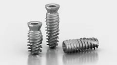
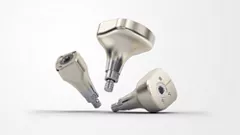
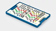
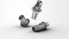
A fully guided digital workflow was selected, incorporating the complete Anthogyr surgical and restorative ecosystem.¹ The Axiom® X3S implant was chosen to enhance primary stability in low-density regenerated bone through its tapered macro-design and aggressive thread geometry, while preserving the same prosthetic trilobe connection as the Axiom® X3 system. Following implant placement, an anatomically shaped, scannable healing abutment, HealFit® SH, was used to guide soft tissue maturation and support a simplified digital workflow, followed by seamless integration with the X-Base® Ti-base restorative solution.²˒³˒⁵
Together, these technologies streamlined the clinical workflow, minimised chair-time, preserved the maturing peri-implant tissues and enhanced communication between clinician and laboratory, improving overall predictability and patient experience.
Fig 1: Initial clinical presentation showing fractured UR2 with retained root remnants and severe upper and lower crowding.
Fig 2: Pre-operative OPG xray demonstrating limited mesio-distal space, compromising ideal implant positioning.
Treatment Plan.
A prosthetic-driven, staged multidisciplinary approach was planned to overcome anatomical constraints and achieve predictable stability in regenerated low-density bone. The strategy combined space optimisation and ridge reconstruction to enable prosthetically guided implant placement, followed by delayed implantation using an adapted protocol to maximise primary stability in D3–D4 bone. The rehabilitation was completed through a streamlined digital workflow designed to preserve peri-implant tissue architecture and ensure efficient, aesthetic integration.
Surgical procedure.
Surgical management was performed in a staged approach to optimise site conditions prior to implantation. Following atraumatic extraction of the non-restorable UR2, initial healing was combined with orthodontic optimisation (ClearCorrect®). Guided bone regeneration (GBR) was subsequently carried out using a combination of xenograft (Cerabone plus®, Botiss) and autogenous bone, together with soft tissue augmentation using an acellular collagen matrix (Mucoderm®, Botiss) to reconstruct ridge volume and enhance buccal tissue thickness in the aesthetic zone (Fig 3-4).
After an 8‑month healing period, implant placement was performed using a fully guided, prosthetic-driven protocol. Digital planning (SMOP®) ensured precise three-dimensional positioning in accordance with restorative objectives. Site preparation followed an under-preparation protocol adapted to regenerated low-density (D3–D4) bone, aimed at maximising mechanical engagement (Fig 5).
The Axiom X3S® implant (Ø3.4 × 12 mm) was placed with high primary stability (>45 N·cm), supported by its tapered design and aggressive thread geometry, enabling controlled insertion and optimal anchorage in the grafted site. A transmucosal approach was subsequently adopted, with immediate placement of a HealFit® SH anatomical healing abutment and simultaneous soft tissue optimisation to support emergence profile and peri-implant tissue maturation (Fig 6-10).
This surgical protocol enabled predictable implant placement in a regenerated low-density environment, while simplifying the workflow and establishing stable conditions for prosthetic rehabilitation.
Fig 3: Post-orthodontic situation. Although alignment improved, residual anatomical limitations and insufficient ridge volume remained, indicating the need for hard and soft tissue augmentation.
Fig 4: Guided bone regeneration using Cerabone plus® xenograft and Jason® collagen membrane and simultaneous soft tissue augmentation using Mucoderm® to enhance buccal tissue thickness and improve long-term soft tissue stability in the aesthetic zone.
Fig 5: Radiographic situation after 8 months of healing. Improved ridge contour allowed for prosthetic-driven implant planning.
Fig 5: Digital implant and Healfit-SH® planning on Smop® (Swissmeda)
Fig 7: Full-thickness flap with palatal extension to allow roll-flap soft tissue enhancement. A tooth supported SMOP® surgical guide was used to ensure accurate prosthetic-driven implant positioning.
Fig 8: Placement of Axiom X3S® implant (Ø3.4 × 12 mm). Implant positioned approximately 1.5 mm subcrestally to support optimal emergence profile and long-term soft tissue stability, achieving high primary stability >45N.cm. The internal trilobe connection orientated buccally to facilitate the correct positioning of the Healfit-SH®.
Fig 9: Buccal roll-flap technique performed to increase buccal soft tissue thickness and support the gingival margin in the aesthetic zone.
Fig 10: Healfit-SH® placement and radiographic verification. Combined suturing using 4/0 resorbable PGA resorba® sutures for internal stabilisation and non-resorbable 5/0 Prolene® sutures for precise papilla adaptation around the healing abutment.


Feel the grip. Master stability.
Discover Anthogyr's new implant solution designed to enhanced anchorage in in demanding clinical situations.
Prosthetic Stage.
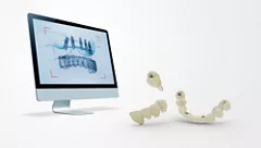
Prosthetic rehabilitation was performed using a fully digital workflow, beginning with intraoral scanning carried out with the HealFit® SH abutment maintained in situ, allowing accurate capture of the conditioned soft tissue profile without the need for abutment removal or additional scan bodies. A CAD/CAM screw-retained implant crown was fabricated on an X‑Base® titanium base, ensuring prosthetic stability and continuity with the Axiom X3S® system (Fig 11-13).
Final aesthetic rehabilitation also included two lithium disilicate (E.max®) veneers on UR1 and UR3, together with a three-unit E.max® bridge on the left side to close the residual spacing associated with the congenitally missing UL2. The conventional restorative treatment and the previous aligner treatment was completed by Dr Konstantinos Karatzioulas (Fig 14-16).
The combination of a scannable healing abutment and a fully digital workflow enabled preservation of peri-implant soft tissue architecture, reduced clinical steps, and improved communication between clinician and laboratory, contributing to a predictable and efficient restorative outcome.
Prosthetic Stage.
Long-term supportive periodontal and peri-implant maintenance program with regular clinical and radiographic review and hygiene appointments.
Fig 11: Three-week follow-up. Sutures removed, demonstrating favourable healing and early maturation of peri-implant soft tissues around the anatomic healing abutment. Healfit-SH® positioned >1.5 mm above soft tissue according to protocol.
Fig 12: Teeth UR1 and UR3 were prepared for E.max® veneers, while the left side was prepared for a three-unit E.max® bridge to close the residual spacing associated with the congenitally missing UL2. The HealFit-SH® scannable anatomical healing abutment allowed simultaneous intraoral scanning without removal of the abutment or placement of a separate scan body, preserving the conditioned soft tissue profile and simplifying the fully digital workflow.
Fig 13: Provisionalisation phase using a pontic positioned over the HealFit-SH® abutment. The patient’s low smile line allowed a simplified temporary approach while preserving the conditioned peri-implant soft tissues. Throughout the aligner phase, aesthetics were maintained using a composite tooth incorporated within the aligners, ensuring the patient was never left with a visible anterior gap.
Fig 14: Soft tissue contour at the time of definitive restoration delivery following removal of the HealFit-SH®, demonstrating stable peri-implant tissues, enhanced buccal soft tissue volume and a well-defined emergence profile.
Fig 15: Definitive restorations delivered, including a screw-retained layered zirconia implant crown on an X-Base® titanium base at UR2.
Fig 16: Final periapical radiograph showing complete seating of the definitive crown and stable peri-implant bone levels at delivery.
Conclusion.
This case demonstrates that a prosthetic-driven multidisciplinary approach enables predictable rehabilitation in the aesthetic zone despite anatomical constraints and regenerated low-density bone conditions. The use of the Axiom X3S® implant supported reliable primary stability and simplified clinical execution, while integrating seamlessly within a digital workflow to preserve peri-implant tissue architecture and achieve a stable, aesthetic outcome.
Clinical Takeway.
In regenerated low-density bone, a multidisciplinary and guided approach using Axiom® X3S enables reliable primary stability and predictable aesthetic outcomes in the anterior zone.
References.
- Schubert O, Schweiger J, Stimmelmayr M. Digital implant planning and guided implant surgery – workflow and reliability. Br Dent J. 2019;226:101–108.
- Kim JJ, Park JH, Choi JH. Accuracy of implant impression techniques with a scannable healing abutment: an in vitro study. J Prosthet Dent. 2021;125(6):903–909.
- Alqahtani F, Mahesh L, Reddy M. Custom anatomic healing abutments for peri-implant soft tissue contouring: a clinical review. J Indian Prosthodont Soc. 2016;16(4):389–394.
- Trisi P, Berardini M, Falco A. Osseodensification: an innovative technique with manifold gains. J Clin Med. 2023;12(22):7046.
- Sanz-Martín I, Martin C, Romanos G. A novel coded healing abutment for a simplified digital workflow: clinical and laboratory validation. J Prosthet Dent. 2024;132(4):560–567.