






 International – English



Transcrestal Sinus Floor Elevation With Simultaneous Placement Of An Anthogyr Axiom® X3S Implant And Restoration With 3D-Printed Crown On An X-BASE® Abutment

Dr. Giuliano Fragola
Spain
Qualified Specialist in Oral Surgery
- DDS, MS, PhD Madrid Complutense University 1995
- Master science in implant dentistry 1996
- Quintessence Spanish translator and reviewer
- ITI Senior Fellow – Anthogyr national and international speaker
- CEO Immersive Dental Experiences IDE. Virtual Reality dentistry apps
- CEO Kyndora Software. Customized dental software
- Dental surgeon. Private practice Madrid, Spain
Introduction.
Implant placement in the posterior maxilla is often challenged by reduced bone height and low bone density due to alveolar resorption and sinus pneumatization after tooth loss [1]. In such cases, sinus floor elevation is required to enable implant rehabilitation [2].
The transcrestal (indirect) sinus lift approach is a minimally invasive technique indicated when residual bone height is moderate, allowing simultaneous implant placement while reducing surgical morbidity and treatment time [3]. When combined with bone grafting and implants designed for soft bone, this approach supports predictable primary stability and successful osseointegration [4].
Initial situation.
A 47-year-old male patient attended our clinic requesting evaluation for the replacement of a missing molar. The patient reported that the tooth had previously undergone root canal treatment and subsequently fractured, leading to its extraction eight years earlier.
From a general medical perspective, the patient was healthy, with no relevant systemic diseases. Oral hygiene was adequate, and both plaque and bleeding indices were within normal limits, so no complementary periodontal or hygiene treatment was required before implant therapy.
A clinical intraoral examination was performed. The restorative space remained stable, with no displacement or mobility of the adjacent teeth and no extrusion of the opposing dentition.
A CBCT scan was taken to assess the available bone height and width. The missing tooth was in a subsinusal position (16), with marked resorption of the alveolar crest and pneumatization of the right maxillary sinus (Fig 1).
Fig. 1–2 — CBCT evaluation of 16 and implant planning.
Treatment Plan.
After radiological evaluation and digital planning in CoDiagnostix software (Fig 2a-c), the treatment plan consisted of placing a dental implant combined with an atraumatic transcrestal sinus floor elevation and simultaneous transcrestal grafting.
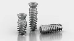
An Anthogyr Axiom® X3S 4.6/8 mm implant (AT46080) was selected for this case. The reason for choosing this implant was the low bone density observed in the posterior maxilla. The macro design of the Axiom® X3S provides apical engagement through its active apical threads, while its parallel body walls and specific implant channels allow bone compaction within the implant bed. The non-cutting edges of these channels are designed to condense rather than remove bone, helping to improve primary stability in low-density bone.
Surgical procedure.
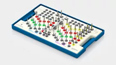
The implant was placed using a two-stage surgical approach with submerged healing. The surgical protocol consisted of step-by-step drilling with drill stops up to 6 mm (Fig 3a-c), followed by transcrestal sinus floor elevation using osteotomes according to the Summers technique. XenoOss Plus xenograft material was introduced through the implant bed to achieve and stabilize the elevation of the sinus membrane, creating a grafted area between the implant apex and the elevated Schneiderian membrane, as described in the Summers technique (Fig 3d).
Fig 3-7: Freehand site preparation followed by a progressive minimal drilling protocol using a 2.4 mm drill to a depth of 4 mm. Atraumatic transcrestal sinus floor elevation was performed, and xenograft material (XenoOss Plus) was introduced. The Axiom® X3S implant (AT46080) was then placed, achieving apical engagement for primary stability (45N.cm). Controlled insertion was carried out with minimal bone compression. Final verification of implant position was performed prior to suturing.
After an osseointegration period of four months, a minimal crestal incision was performed to place a healing abutment, reference OPHSF620, to condition the peri-implant soft tissues before the prosthetic phase (Fig 8-11).
Fig 8-11: Four months follow up radiograph and healing.


Feel the grip. Master stability.
Discover Anthogyr's new implant solution designed to enhanced anchorage in in demanding clinical situations.
Prosthetic Procedure.
After 15 days of soft tissue healing, an intraoral scan was carried out using the Straumann SIRIOS X3 scanner with the crown and bridge protocol. First, the upper and lower arches were scanned, together with the occlusal relationship, while maintaining the healing abutment in position in the upper arch.
The area corresponding to the healing abutment was then identified and digitally trimmed from the scan. The healing abutment was removed, and the peri-implant soft tissue was scanned in high definition to accurately capture the emergence profile. A 152-27-MDT scanbody was then placed and scanned.
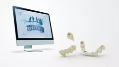
Once the file had been processed by the SIRIOS software, the case was transferred directly to the MIDAS Smart Design system. The scanbody was identified, and a titanium base was selected, as the final restoration was planned as a cement-retained crown on an X-Base®.
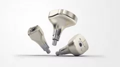
The selected X-Base® was the OPFLEX 414, with a 4 mm emergence profile and a 1.5 mm height. Following the digital design steps in MIDAS, the definitive crown was designed. A Crown Permanent A3 cartridge was then selected and inserted into the MIDAS printer.
The crown was 3D printed according to the manufacturer’s protocol, followed by the corresponding post-processing, curing and characterization procedures. Subsequently, the X-Base® was cemented extraorally to the crown. The margins were carefully polished and inspected to ensure that no residual cement remained.
Finally, the restoration was placed intraorally, the screw access was managed according to the clinical protocol, and the occlusion was checked and adjusted to achieve a stable and functional result.
Fig 12-16: IOS, Midas Smart Design and chairside printing.
Final Situation.
Once polished and characterized, the final restoration was placed according to the recommended insertion torque for the prosthetic XBase abutment (25 Ncm), and the occlusion was carefully checked. The patient was instructed on the appropriate oral hygiene measures and advised to return for a follow-up appointment after 15 days and subsequently at 6 months.
Fig 17-18: final restoration after characterizaton and placement.
Conclusion.
This clinical case shows that minimally invasive transcrestal sinus floor elevation with simultaneous implant placement can be successfully performed in a posterior maxilla with reduced bone height and low bone density. The Axiom X3S implant allowed apical engagement and bone condensation, supporting primary stability in this indication. The combination of this surgical approach with digital workflow in the prosthetic phase enabled an efficient and controlled restorative process.
Clinical Takeway.
Axiom® X3S delivers reliable primary stability in soft bone, enabling safe, minimally invasive sinus lift with simultaneous placement.
References.
- Lee JE, Jin SH, Ko Y, Park JB. Evaluation of anatomical considerations in the posterior maxillae for sinus augmentation. World J Clin Cases. 2014 Nov 16;2(11):683-8. doi: 10.12998/wjcc.v2.i11.663. PMID: 25405192; PMCID: PMC4233423.
- Kim YK, Ku JK. Sinus membrane elevation and implant placement. J Korean Assoc Oral Maxillofac Surg. 2020 Aug 31;46(4):292-298. doi: 10.5125/jkaoms.2020.46.4.292. PMID: 32855378; PMCID: PMC7469962.
- Franceschetti, G., et al. “Transcrestal Maxillary Sinus Floor Elevation with Bone Substitutes. A Prospective Case-Control Study.” Proceedings Book Research Session “Henry M. Goldman”, Research Centre for the Study of Periodontal and Peri-implant Diseases, University of Ferrara, Ferrara, Italy.
- Firlej M, Huwais S, Neiva R. Crestal Sinus Lift With Osseodensification in Severely Atrophic Maxilla: Case Series With Long-Term Follow-up. Compend Contin Educ Dent. 2025 Jan-Feb;47(1):e9-e12. PMID: 41627999.