






 International – English



Predictable Primary Stability in an Immediate Post‑Extraction Socket Using Axiom® X3S and Fully Guided Surgery

Dr. Levs GAVRILOVS
Spilsby, United Kingdom
Doctor of dental surgery specialized
in implant and restorative dentistry
- Postgraduate from the British Academy of Implant
and Restorative Dentistry, Leeds (UK) - Postgraduate Advanced Implant Dentistry, Milan (IT)
- Postgraduate Advanced Aesthetic Dentistry, London (UK)
- Private practice in Spilsby (UK)
- International speaker
Introduction.
Immediate implant placement in post-extraction sockets remains a technically demanding indication because primary stability and implant positioning must be controlled within a compromised anatomical environment. Success depends on achieving mechanical anchorage in native bone while maintaining favorable biologic conditions for healing (1-3).
This case report describes immediate implant placement in a mandibular premolar socket using a fully digital guided workflow and the Axiom® X3S implant system. The aim is to present a stability-driven, risk-controlled approach in which primary stability was obtained without grafting and loading was deliberately delayed to favour osseointegration.
Initial situation.
A 60‑year‑old non‑smoking patient presented with a fractured and unrestorable retained root of the lower right first premolar (LR4). Clinical examination confirmed the indication for extraction, and comprehensive pre‑operative diagnostics were performed, including CBCT imaging, intraoral scanning, and clinical photography. The CBCT analysis was used to assess socket anatomy, residual bone availability, and prosthetic constraints, providing the basis for prosthetically driven digital planning (Fig 1-4 ).
Fig 1-4. Initial situation and diagnostic assessment. Preoperative clinical photographs and CBCT analysis showing a fractured, non-restorable retained root at site LR4. Radiographic evaluation illustrates socket anatomy, residual bone availability, and prosthetic constraints guiding treatment planning.
Treatment plan.
Following clinical and radiographic evaluation, the treatment plan consisted of atraumatic extraction of the fractured and unrestorable retained root at site LR4, followed by immediate implant placement using a fully guided digital workflow.
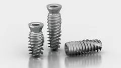
The Axiom® X3S implant was selected to optimize primary stability in the post‑extraction socket, with simultaneous guided bone regeneration planned to support hard and soft tissue healing. A delayed loading protocol was chosen to prioritize biological integration, with final restoration planned using a CAD/CAM‑fabricated customized abutment and a full‑zirconia crown.
Alternative treatment options, including delayed implant placement, were discussed with the patient, and informed consent was obtained.


Feel the grip. Master stability.
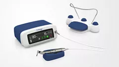
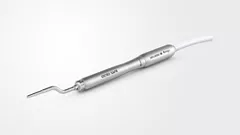
Discover Anthogyr's new implant solution designed to enhanced anchorage in in demanding clinical situations.
Surgical procedure.
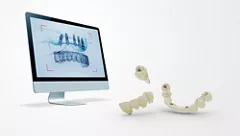
Digital treatment planning was carried out using SMOP software by merging CBCT and intraoral scan data to define implant position, axis, and depth. A fully guided surgical template was designed and 3D‑printed prior to surgery.
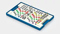
The retained root was extracted atraumatically with careful preservation of the socket walls, followed by meticulous debridement and irrigation Implant placement was performed using the Anthogyr Integral Guided Surgery Kit, and the Axiom® X3S implant achieved a primary stability of 35 Ncm (Fig 5-9).
A healing abutment was placed at the time of surgery, and no immediate provisionalization was performed (Fig 10).
Simultaneous guided bone regeneration was carried out for gap management using Maxgraft allograft material, in combination with Mucoderm as a soft‑tissue substitute serving as a barrier membrane to support both hard and soft tissue healing.
Fig 5-10. Atraumatic extraction followed by fully guided implant placement of an Axiom® X3S implant in the socket (35 N.cm primary stability), with healing screw placement and no immediate provisionalization (delayed loading protocol).
Prosthetic restoration.
After an uneventful healing period of twelve weeks, clinical evaluation confirmed successful osseointegration (Fig 11-13 ).
Fig 11-13. Healing and osseointegration phase. Twelve-week postoperative evaluation demonstrating uneventful healing and successful osseointegration, with stable peri-implant soft tissues.
A new intraoral scan was performed to initiate the prosthetic phase.
A CAD/CAM‑customized abutment was fabricated, followed by delivery of a definitive full‑zirconia crown, providing stable occlusal function and appropriate integration within the posterior mandibular region (Fig 14-22).
Fig 14-22. Prosthetic workflow and final restoration. Digital impression and CAD/CAM fabrication of a customized abutment followed by delivery of a full zirconia crown. Final clinical views show functional integration and harmonious adaptation within the mandibular arch.
Final situation.
At the completion of treatment, the implant‑supported restoration demonstrated stable peri‑implant tissues, satisfactory functional integration, and harmonious adaptation within the dental arch. The patient reported a high level of satisfaction in terms of comfort and function, and no surgical or prosthetic complications were observed throughout the treatment course.
Conclusion.
This case demonstrates that predictable primary stability can be achieved in an immediate post‑extraction mandibular socket when implant selection, planning, and execution are aligned with a stability‑driven clinical strategy. The Axiom® X3S implant provided reliable mechanical anchorage at placement without reliance on grafted bone for stability, while the fully digital, guided workflow enabled controlled implant positioning in a limited anatomical environment. Despite achieving adequate insertion torque, loading was intentionally delayed, reflecting a biology‑first approach, particularly in the presence of simultaneous bone regeneration. Overall, this case highlights how implant macro‑design, guided surgery, and disciplined clinical decision‑making can be combined to manage uncertainty and achieve predictable outcomes in immediate implant indication.
Clinical Takeaway.
A stability-driven approach combining fully guided surgery and Axiom X3S enables predictable primary stability in immediate post-extraction sockets, even without relying on grafted bone for anchorage.
References.
Giannini, M., et al. (2023). Primary stability of implant placement and loading related to placement‑timing strategies. Journal of Clinical Medicine, 12 (12), 4112. https://doi.org/10.3390/jcm12124112
Pozzi, A., et al. (2022). Does guided level (fully or partially) influence implant placement accuracy at post‑extraction sockets and healed sites? An in vitro study. Clinical Oral Implants Research, 33 (11), 1021–1032. https://doi.org/10.1111/clr.13998
Dentistry, P. (2015). Guidelines for immediate vs delayed dental implant placement in the esthetic zone. Pocket Dentistry
Carpentieri, J., & Greenstein, G. (2015). Guidelines for immediate vs. delayed dental implant placement in the esthetic zone. Compendium of Continuing Education in Dentistry.