


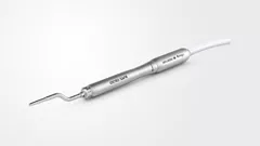

A Sealing Socket Abutment (SSA)
for a wide implant designed
for single molar replacement.

Dr. Sami EL MASRI
Qualified Specialist in Oral Surgery
Belgium
- Masters in Dentistry, University of Louvain, BE
- Specialized Master of Prosthetic Rehabilitation, University of Louvain, BE
- Specialized Master in Oral Surgery and Implantology, University of Liège, BE
- Head Chief of Clinic Dental ME, Louvain la Neuve, BE
- Associated Instructor for Education by Dental ME with Prof. Eric Rompen
and Dr. Martin Désiron
Case introduction
The architecture of molars is intricate; the orientation and positioning of the implant is influenced by the root's location and the socket's structure during immediate implantation surgical techniques. The large socket size facilitates the positioning of wide-diameter implants within the inter-radicular bone at the centre of the multi-rooted socket, while the implant reduces the size of adjacent voids, thereby decreasing the requirement for grafting materials. Application of ultra-wide diameter implants in immediate molar replacement is widely accepted and exhibit minimal bone loss and maintain stable soft tissue conditions over a period of 7 years[1-2].
Conventional healing abutment placement is not appropriate for these broad extraction sockets, which are difficult to seal without mucoperiosteal flap reflection and the crestal socket shape. The use of surgeon-fabricated screw-retained temporary restorations is advantageous for improving gingival contours and achieving desirable aesthetics [3a].
Custom immediate posterior healing abutments provide support for local anatomy during osseointegration and allow direct access to the implant during the restorative phase.
Post-osseointegration, the result is an anatomical representation that mimics the growing profile of real teeth, allowing precise laboratory replication of the final abutment.
This method provides all the advantages of implant provisionalization [3a].
This clinical case illustrates the treatment of a molar by immediate extraction and implantation followed by placement of a sealing socket abutment (SSA).
Initial situation
The patient was diagnosed by 3D CT scan and radiograph during a routine control of a classic first molar. The clinical signs necessitated tooth extraction followed by immediate implantation, socket gap filling and placement of a sealing socket abutment to support anatomical structures.
1. Radiograph of initial situation.

1
2a. 2b. Initial intraoral view of the molar.

2a

2b
3a. 3b. 3c. 3D volumetric diagnosis and planning of implant therapy.

3a

3b

3c
4. Radiograph exhibiting a suitable septum for immediate implant placement.

4
Surgical procedure
5. Occlusal view after atraumatic tooth extraction.

5
6. Immediate implant placement Ø 6.4mm Axiom® X3, 4 mm deep from the gum margin in the bone furcation for mucointegration.

6
7. Filling gaps with xenograft material
for socket preservation.

7
8. Occlusal view of the composite socket sealing with a 3mm provisional titanium retained SSA as fabricated by Gary Finelle.

8

Curious about Axiom X3® profile?
Axiom X3® dental implant is a unique design for controlled insertion and optimal stability in a wide range of indications, with maximum bone preservation regardless
of density.
9. Intra oral view with the SSA in place.

9
10a. 10b. 10c. 3D scans and radiograph control of the implant![]() placement.
placement.

10a

10b

10c
Prosthetic restoration
11a. 11b. Radiographs of the SSA in place and scan body for digital impressions.

11a

11b
12a. 12b. 12c. 12d. Digital files after intra-oral scanning showing the gingival contouring for the prosthetic phase 4 months post implant placement.

12a

12b

12c

12d
Final situation
13. Peri-apical radiograph depicting final prosthesis.

13
14a. 14b. Radiograph of 2 years follow-up.

14a

14b
Conclusion
The wide diameter selected from the Axiom® X3 implant range allowed us to treat this basic molar without large grafts. Patient care was optimal, and follow-ups show stability of solution. For immediate molar replacement, the wide diameter implant increases engagement and contact with the inter-radicular bone and socket walls, which would be limited with a narrower implant.
References
1. Wadhwa P, Kim S-K, Kim H-J, Lim H-K, Jia Q, Jiang H-B, et al. A Six-Year Prospective Comparative Study of Wide and Standard Diameter Implants in the Maxillary and Mandibular Posterior Area. Medicina (Kaunas) 2021. 57(10):
2. Mustakim KR, Eo MY, Lee JY, Myoung H, Seo MH, Kim SM. Guidance and rationale for the immediate implant placement in the maxillary molar. J Korean Assoc Oral Maxillofac Surg 2023. 49(1): pp. 30–42.
3. Hattingh A, Bruyn HD, Vandeweghe S. A retrospective study on ultra-wide diameter dental implants for immediate molar replacement. Clin Implant Dent Relat Res 2019. 21(5): pp. 879–887.
4. Akin R. A New Concept in Maintaining the Emergence Profile in Immediate Posterior Implant Placement: The Anatomic Harmony Abutment. J Oral Maxillofac Surg 2016. 74(12): pp. 2385–2392.