


Post-extraction implant rehabilitation
using Axiom X3® Tissue Level implants
and digital flow.

Dr. Nuño GIL
Qualified Specialist in Oral Surgery
Burgos, Spain
- Degree in Dentistry from the Alfonso X El Sabio
- Specialist in advanced aesthetics
- Diploma in periodontics and implantology from the Implanteperio Group
- Private practice in Burgos, Spain
Case introduction
A 58-year-old patient, a non-smoker in an optimal state of general health came to our clinic with multiple missing teeth that prevented him from performing a correct masticatory function as well as various other periodontal problems. This impacted negatively on his self-esteem and overall well-being, leading him to consider seeking treatment from a dental professional.
Treatment plan
The patient had not received treatment previously due to odontophobia, so we suggested performing the surgery under intravenous sedation to enable a longer treatment in a single session. Due to the presence of generalized periodontitis, the decision was made to extract all remaining teeth and proceed with restoration using Axiom X3® Tissue Level implants.
This approach helped avoid the need for further intervention at the gingival level through non-submerged healing process.
We utilized implants with standard diameters of ≤ 4.0mm to ensure adequate bone volume around each implant, permitting firm anchorage, primary stability and optimal vascularization.
A screw-retained prosthesis made of monolithic zirconia cemented to a titanium structure was custom-designed and manufactured for the upper jaw. For the lower jaw, a similar approach was taken, but the monolithic zirconia was replaced with resin and an unloading splint. All restorations were carried out using a fully digital workflow.
Initial situation
1. Panoramic radiograph view before extraction of teeth in the mandible and maxilla.
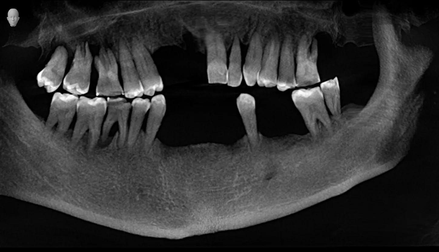
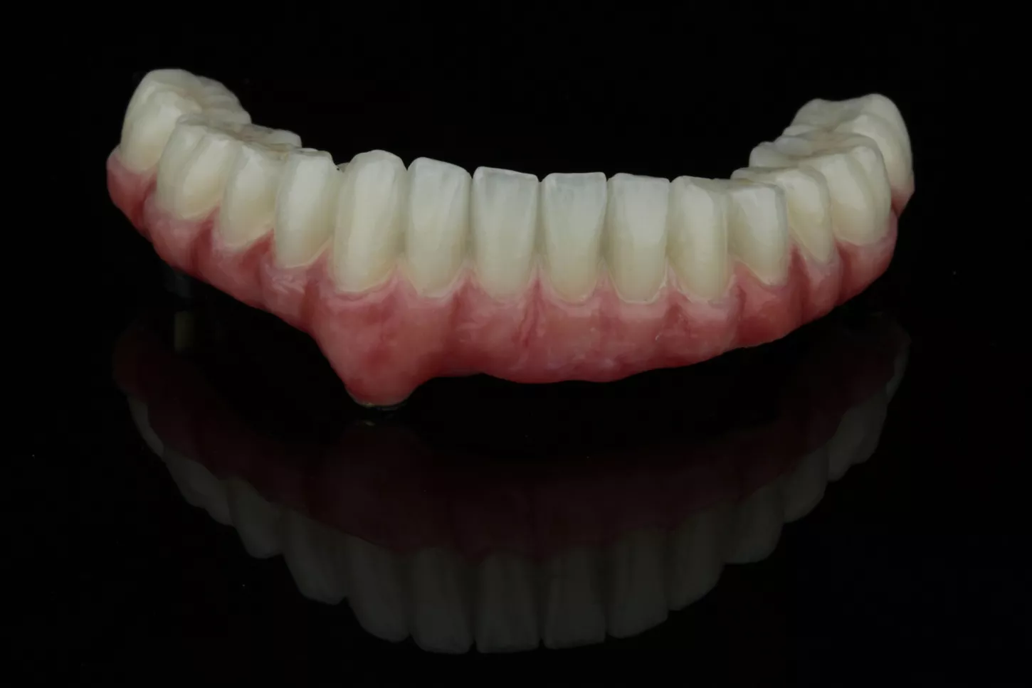
1
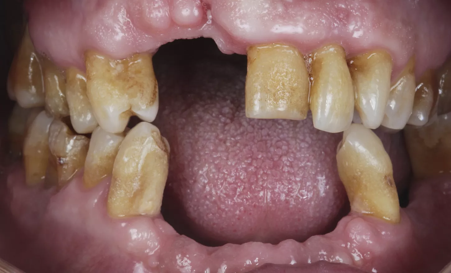
2. Preoperative frontal view showing several missing teeth in both jaws incisor/canine/premolar regions, gingival recession and atrophied alveolar ridges
and remaining carious teeth.
2
Surgical procedure
3a. 3b. Atraumatic extractions of all remaining teeth on maxillary arch to preserve the remaining tissues.
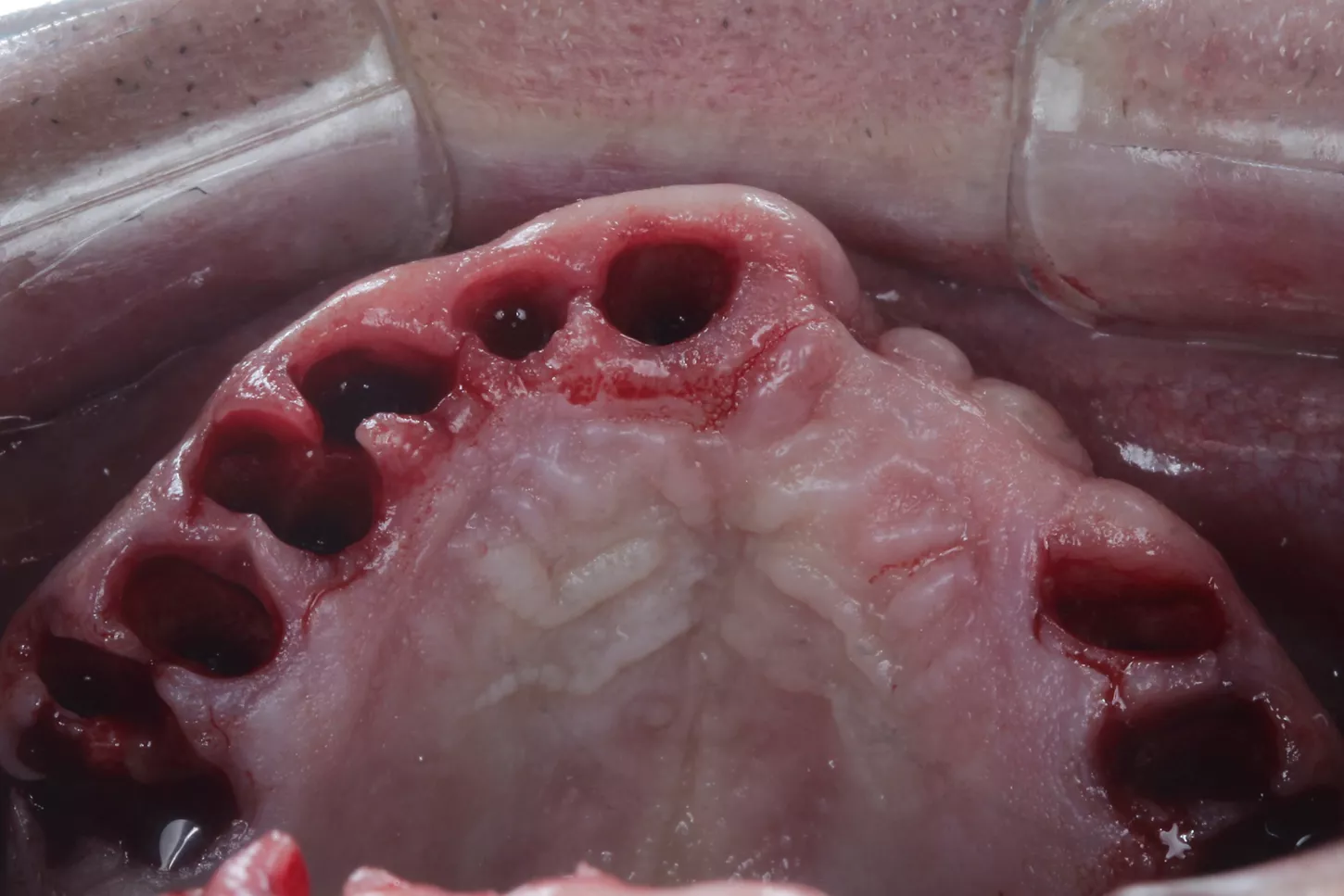
3a
3b
4a. 4b. Prior to implant placement, a full-thickness flap was elevated and bone levelling with a shaping bur was performed.

4a

4b
5a. 5b. The drilling sequence for Axiom X3® Tissue Level implants was according to the bone densities of specific quadrants in order to ensure optimal primary stability of the fixations. The implantation sites in the anterior and posterior zones were determined to be D1/D2 and D2/D3 respectively. The axis and dimensions of the implantation sites were confirmed using appropriate gauges.
A total of 6 Axiom X3 ® Tissue Level N platform implants were utilized on the upper jaw, comprising 3 units of Ø 4.0 x 10mm and 3 units of Ø 4.0 x 12mm, all with a gingival height of 2.5mm.
On the lower jaw, 6 implants were used, including 3 units of Ø 4.0 x 8mm, 2 units of Ø 3.4 x 10mm, and 1 unit of Ø 4.0 x 10mm, all with a gingival height of 2.5mm. The average torque measurement at implantation was recorded to be 60 N.cm.

5a

5b
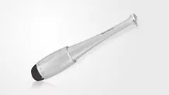
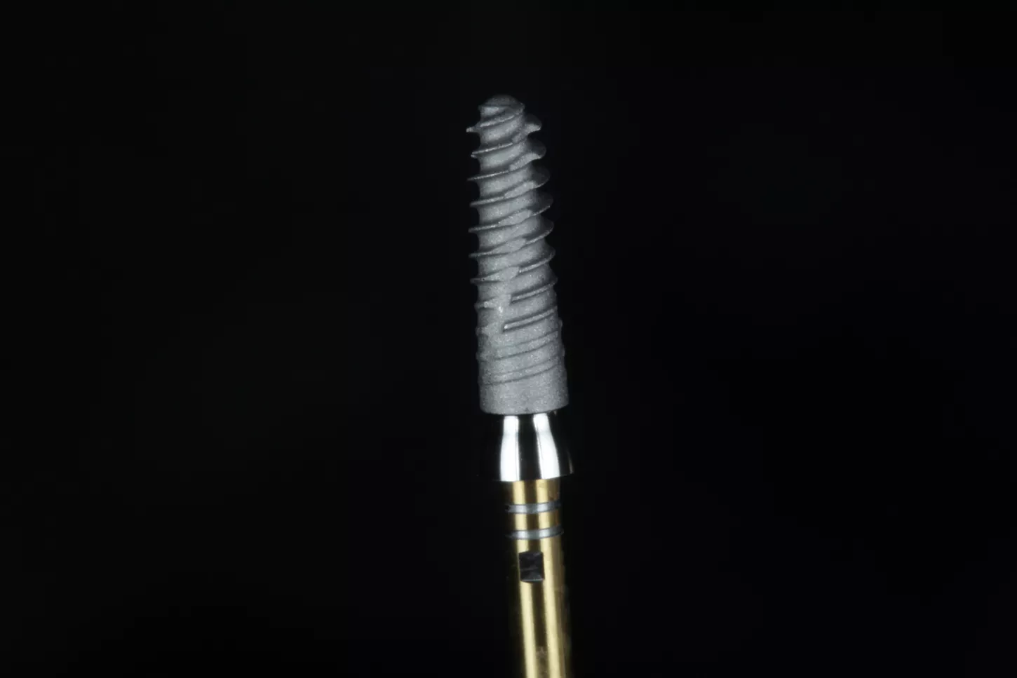
6a. 6b. An example of one of the multiple Axiom X3® Tissue Level N platform implants used in the surgical phase.
6a

6b
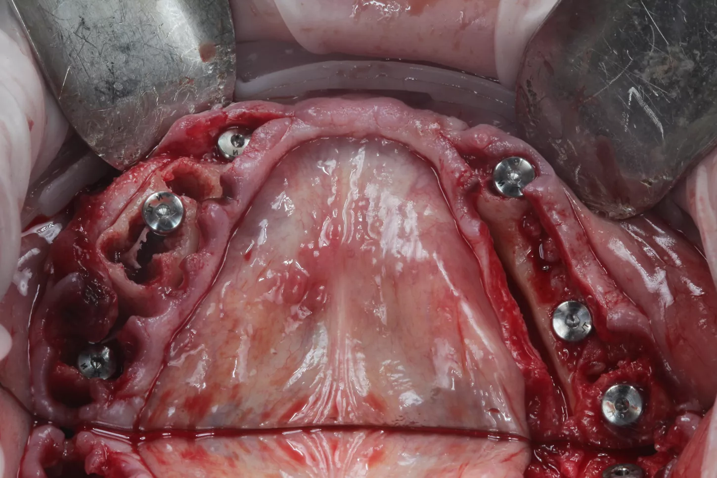
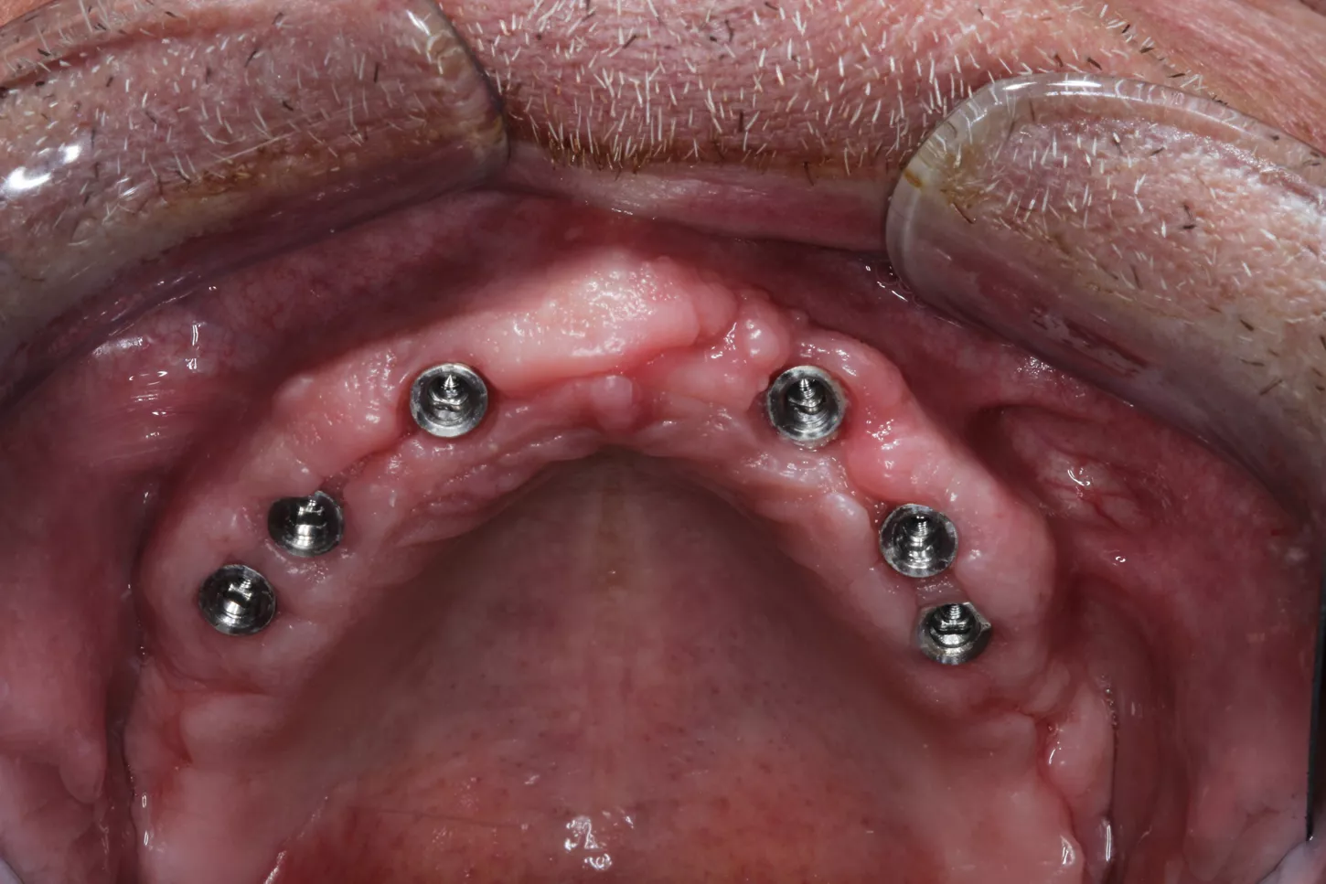
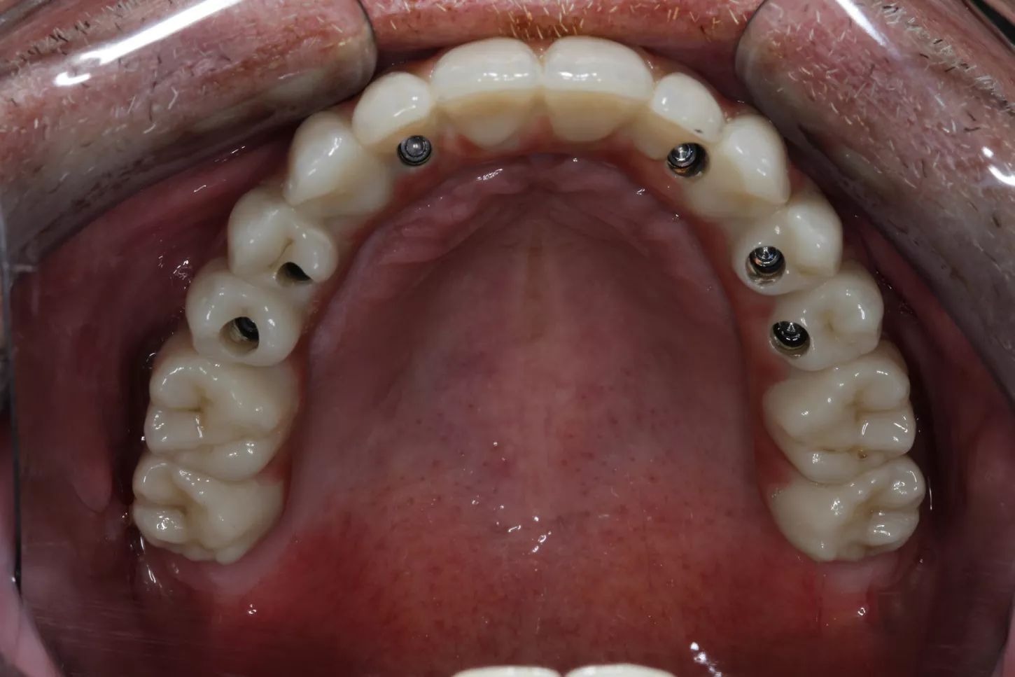
7. Occlusal view with all Axiom X3 ® TL N platform implants placed in the upper jaw. Each implant is fitted with its respective healing cap, which is securely screwed on.

7
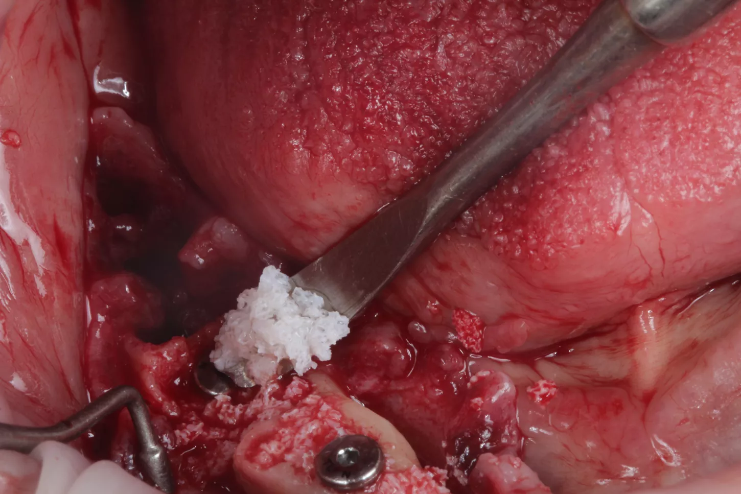
8. The existing gaps were filled with Straumann ® XenoGraft biomaterial to support the bone remodelling processes that occur post extraction.

8
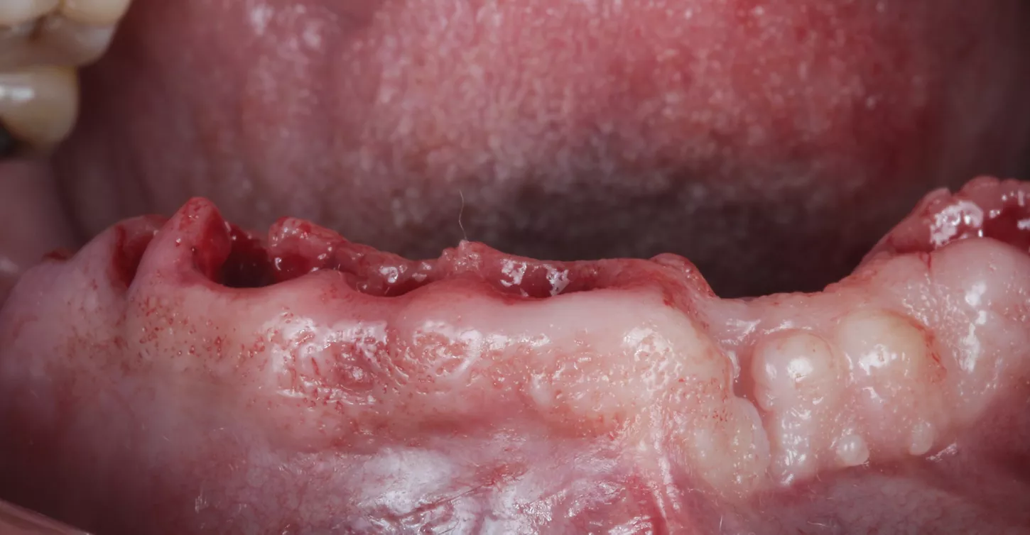
9. Procedures in the lower jaw were similar to those in the upper jaw. This involved carrying out extractions followed by bone regularization.

9
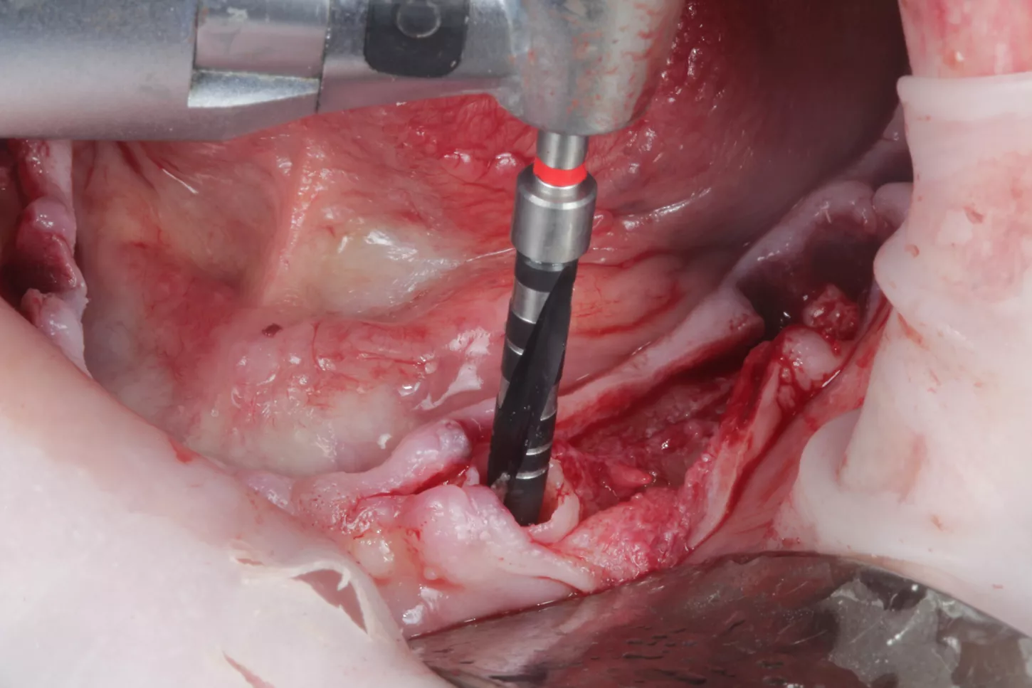
10. Drilling sequence conducted according to the Axiom X3® Tissue Level Ø 3.4 mm x 10mm, gingival height 2.5 mm implant, in this case being mandibular bone of D2, we stopped at the red drill of Ø 2.4/3.0 mm in the Axiom® Surgical Kit.
10
11a. 11b. Placement of the Axiom X3® Tissue Level implants.

11a
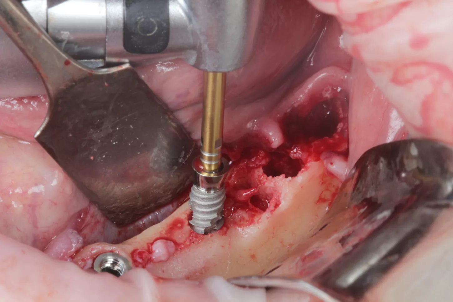
11b

Curious about Axiom X3® profile?
Axiom X3® dental implant is a unique design for controlled insertion and optimal stability in a wide range of indications, with maximum bone preservation regardless
of density.
12a. 12b. Lateral view of simultaneous implant placement in the post extraction sockets in one of the mandibular quadrants.

12a

12b
13. Lateral view of the simultaneous placement of implants in the second mandibular quadrant after the exodontia.
13
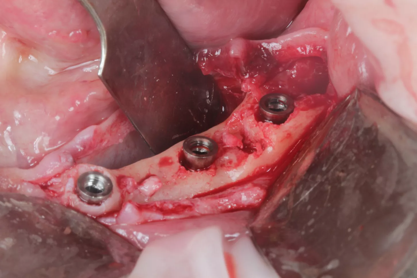
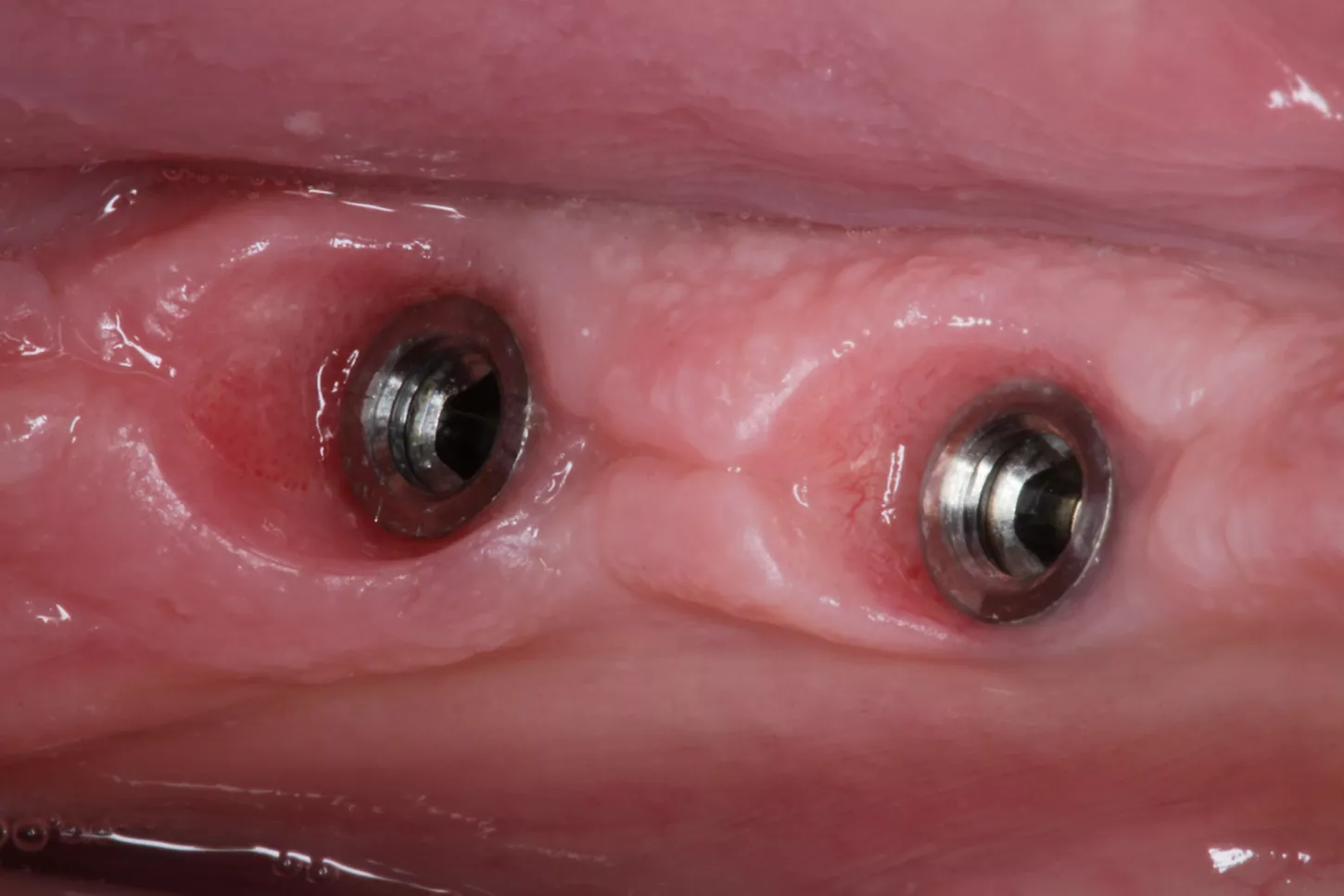
14. Occlusal image of the capped implant placement in the lower jaw.
14
15. The gaps between the implant surface and the internal wall of the socket were filled with Straumann® XenoGraft biomaterial.
15
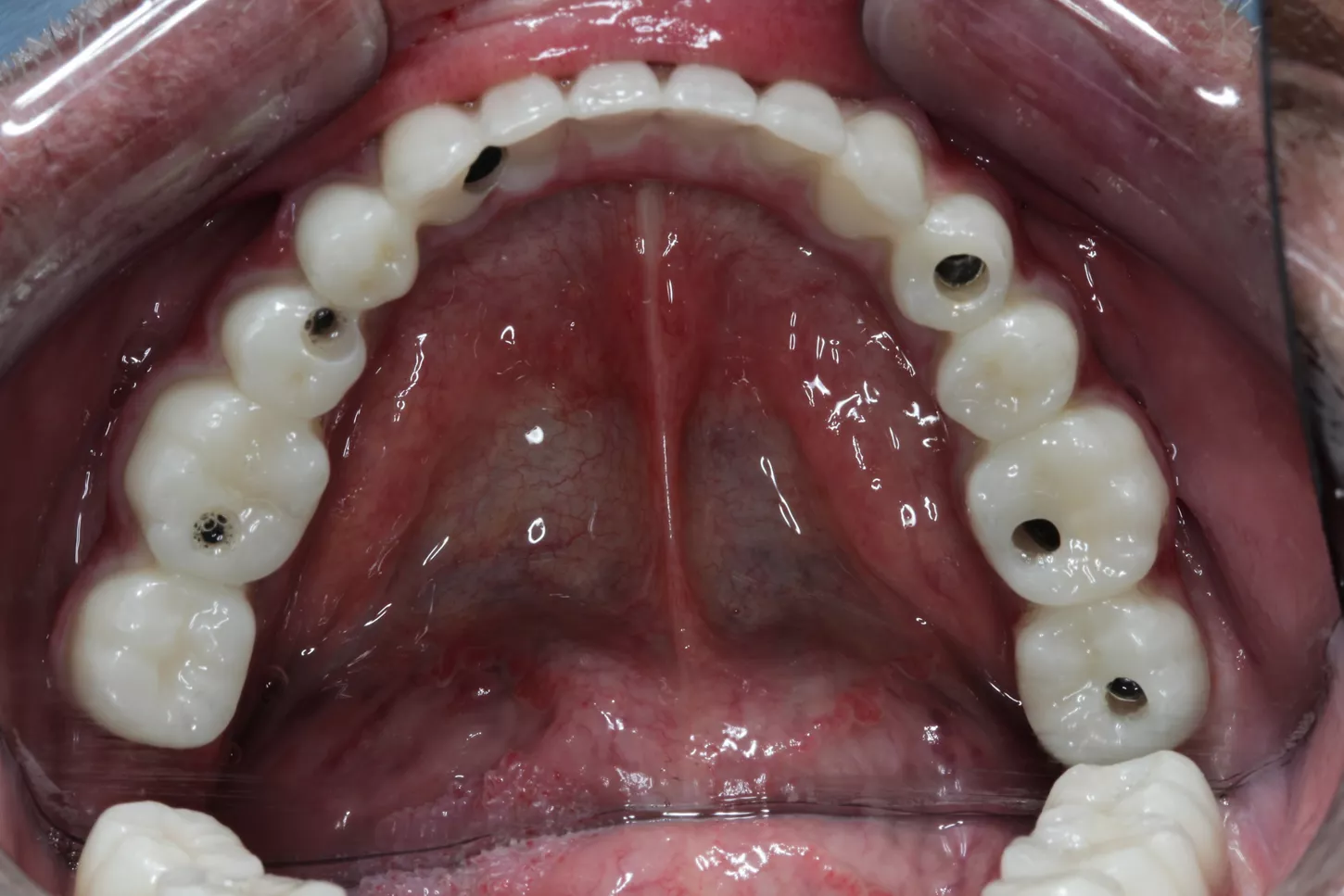
Prosthetic restoration
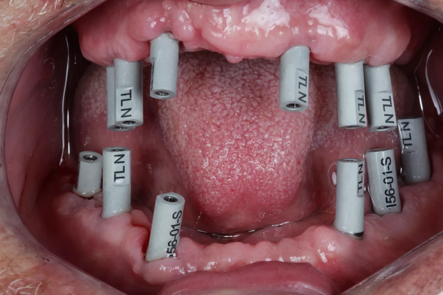
16a. 16b. 16c. The patient received mucosa-supported provisional prosthesis on both jaws. After three months of healing, optimum integration of the soft tissues around the implants neck can be observed. The design of the neck of the Axiom X3® Tissue Level Implant and healing screw permits gingival sculpting.
16a
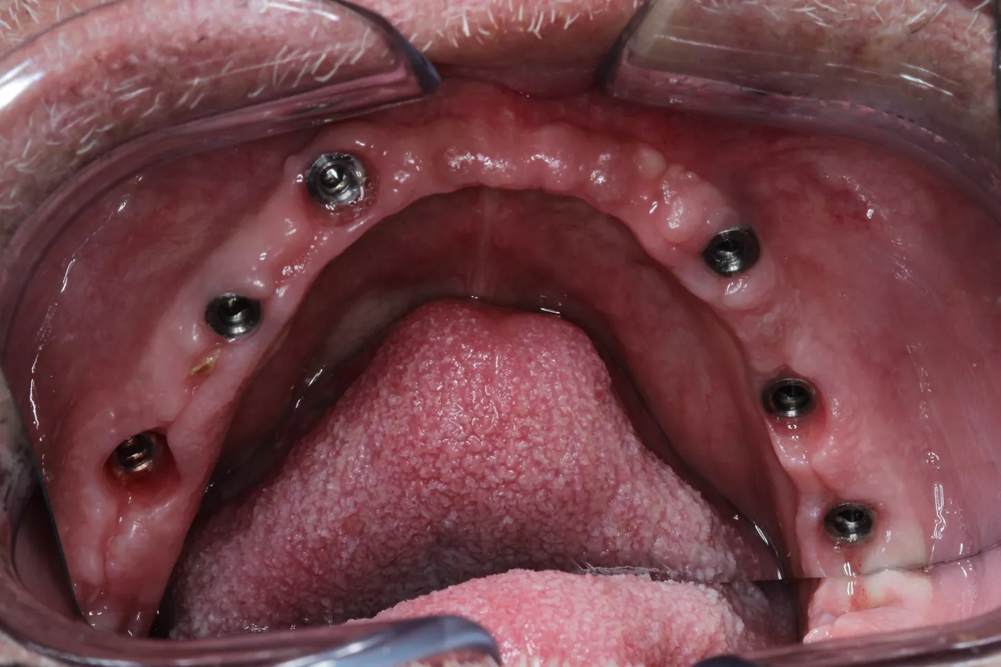
16b
16c
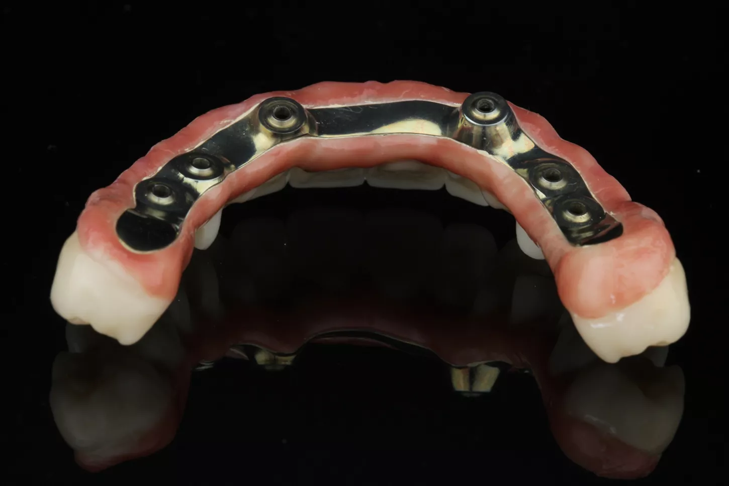
17. Once the designs of our restorations and the various relevant tests had been carried out, we received the prostheses. Both structures (the titanium and monolithic zirconia) were entirely milled in the Simeda® centre and designed by the Corus Garbident laboratory in Vitoria (Spain).
17
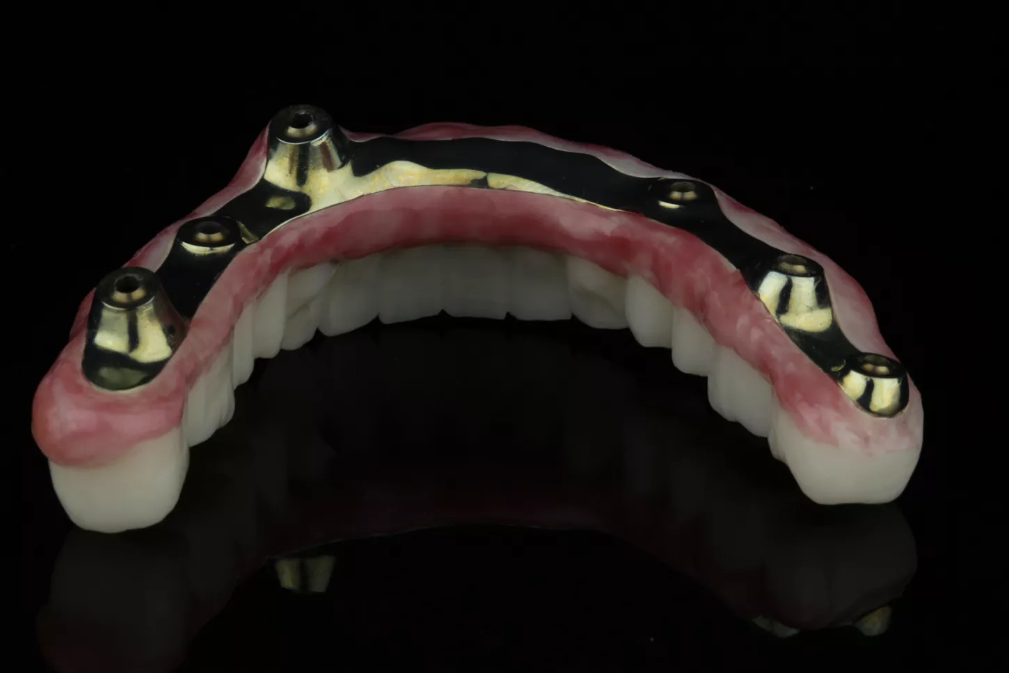
18a. 18b. Images of the upper restoration.
18a
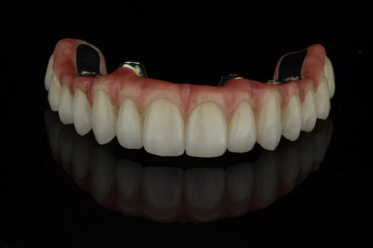
18b
19a. 19b. Images of the lower restoration.
19a
19b
20a. 20b. In this clinical case, and as a novelty, we were able to screw both structures directly to the implants, since, the angled chimneys were incorporated in the titanium structure itself, in this case anodized to improve the final aesthetics.
20a
20b
Final situation
21. Front view of the finished case and placed with both structures screwed in the upper and lower jaw.
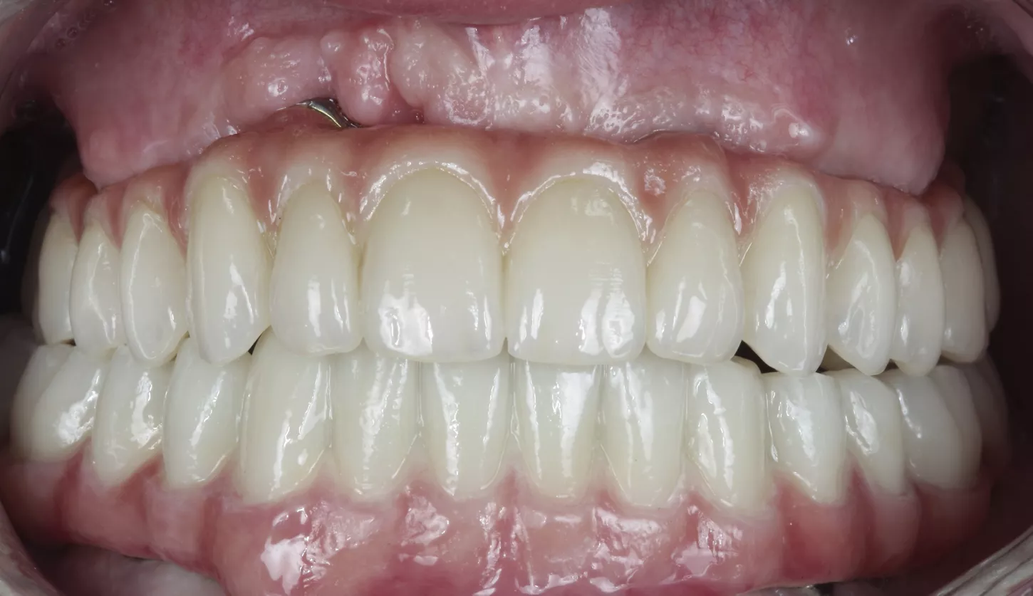
21
22. Post operative radiographs of the implants and with restorations screwed in place.
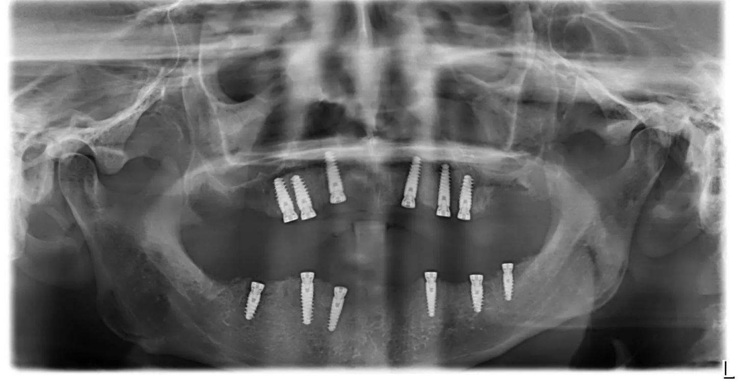
22
Conclusion
The advantages of post-extraction implantology for tissue preservation are well documented [1-4]. In this case, we encountered the challenge of unequal hard and soft tissue volumes and heights in already healed areas versus post-extraction sockets. The possibility to compensate for these deficiencies and allow for tissue remodeling in one surgical act is a great alternative that reduces the need for more complex reconstructive surgeries, decreases patient morbidity, and reduces waiting times for treatment completion allowing for treatment to be completed earlier.
The adaptative drilling protocol of Axiom X3® Tissue Level Implant permitted us to reach appropriate primary stability in different bone types and a favorable remodeling of the soft tissue around the specific implant neck.
References
1. Werbitt MJ, Goldberg PV. The immediate implant: bone preservation and bone regeneration. Int J Periodontics Restorative Dent 1992. 12(3): pp. 206–17.
2. Mura P. Immediate Loading of Tapered Implants Placed in Postextraction Sockets: Retrospective Analysis of the 5‐Year Clinical Outcome. Clin Implant Dent Relat Res 2012. 14(4): pp. 565–574.
3. Ebenezer V, Balakrishnan K, Asir RD, Sragunar B. Immediate placement of endosseous implants into the extraction sockets. J Pharm Bioallied Sci 2015. 7(5): p. 234.
4. Weigl P, Strangio A. The impact of immediately placed and restored single-tooth implants on hard and soft tissues in the anterior maxilla. Eur J Oral Implantol 2016. 9 Suppl 1: pp. S89–106.