


Rehabilitation of a edentulous mandibular arch using Anthogyr mini implant system with Optiloc® connection, an immediate stabilisation solution for removable prostheses.

Dr. Nicolas JUNG
Qualified Specialist in Oral Surgery
Annecy, France
- Practising exclusively in oral surgery and periodontology
- Graduate of the Faculty of Lyon
- Les Aravis Clinic – Annecy
Case introduction
A 70-year-old female patient in good general health but with a history of periodontal disease was referred to find a more durable solution for her mandible. The two remaining canines were mobile (mobility grade 1 for 33 / mobility grade 3 for 43) and supported an unstable Stellite restoration that was causing the patient pain.
Clinical and radiological examination revealed terminal periodontal disease on tooth 43 and advanced periodontal disease on tooth 33. The adjacent bone volume was adequate vertically, but the residual alveolar ridge was thin due to previous extractions (5 mm vestibulo-lingually on average). Periodontal cleaning was performed, and the soft tissues were healthy. Keratinised tissue was present throughout the residual alveolar ridge with a thickness greater than 1 mm.
Treatment plan
In agreement with the treating practitioner (Dr Thomas BLANCHARD) and the patient, it was decided to remove the two canines and consider a complete stabilised solution on 4 Mini implants with Optiloc® connectors. These implants were planned to be distributed across the symphyseal region opposite 32 and 42, as well as mesial to the mental foramen in 34 and 44. This distribution would limit the rocking motion of the future prosthesis [1-2] . A fixed alternative solution using six implants was proposed, but the budget did not allow for this solution to be chosen.
Initial situation
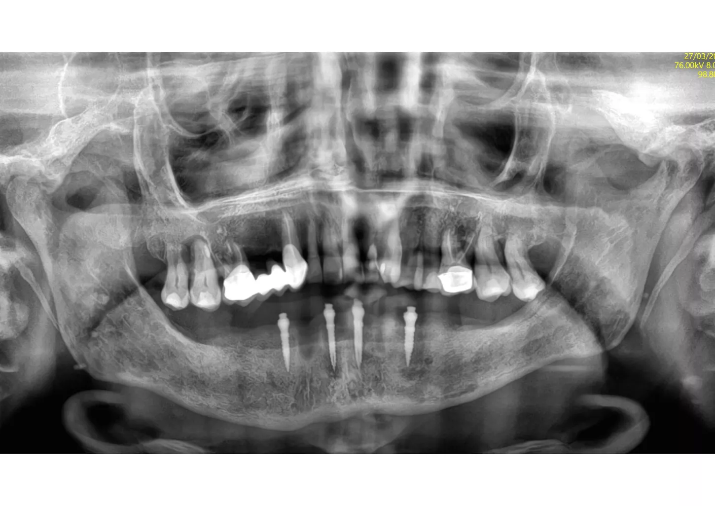
1a. 1b. Pre-operative panoramic X-ray and CBCT showing moderate generalised bone resorption in the mandible, a reduction in vertical dimension in the posterior region and the planned positioning of mini- implants.

1a

1b
2. Intraoral examination.
2a. View of the mouth showing limited gingival exposure.

2a
2b. Situation in the maxilla: the periodontal situation is stable.

2b
2c. Situation in the mandible: thin alveolar crest and residual teeth 33 and 43.

2c
Surgical procedure
3a. Elevation of the full-thickness flap by crestal incision centred on the keratinised and sulcular tissues in 33 and 43.
The canines are preserved to provide an indication of the drilling axis.
Initial drilling in 32 and 42 using the first 1.5 mm drill bit, then positioning of directional indicators to assist with subsequent drilling. Drilling to 12 mm is checked using the graduation on the initial drill bit.

3a

3b
3b. Drilling adjacent to the remaining teeth 33 and 43 using the initial 1.5 mm drill. Depth and axis checked using the anterior indicators and residual canines.
Given the average bone density (D2 to D3), only the initial drill was used to the working length of 12 mm.
3c. Two mini-implants with a diameter of 2.6 mm and a length of 12 mm were placed in positions 32 and 42, first manually and then using a contra-angle. The insertion torque recorded at the end of the procedure was 21 N.cm in 32 and 17 N.cm in 42. The implants were positioned 0.5 mm subcrestal, i.e. with slight burial of the smooth neck.

3c

3d
3d. Two mini-implants with a diameter of 2.6 mm and a length of 12 mm were placed in positions 34 and 44 using the same protocol as for the previous mini-implants. The torque obtained was 27 Ncm in 34 and 37 Ncm in 44.
3e. Canines 33 and 43 were extracted and the burial of the implants was then checked.

3e
3f. 5/0 absorbable sutures were placed around the mini implants, taking care to preserve as much keratinised tissue as possible around the Optiloc attachments.

3f

Curious about our
Mini implant system?
Benefit from a one-piece small diameter dental implant, combined with an integrated Optiloc® prosthetic connection, to offer minimally invasive treatment.
4. Placement of silicone mounting rings, then matrices with white PEEK retention inserts (light retention) on each Optiloc® connector.

4
5. After checking that the matrices did not interfere with the prosthetic intrados and trimming the latter, secured the female parts (matrices) in the mouth using a resin such as Unifast.
Polish off any excess once the resin has set.

5
6. Post-operative X-ray showing the implants in place as planned.
6
Prosthetic restoration
7a. 7b. Post-operative view of the complete removable prosthesis immediately secured to the mini implants with Optiloc connectors.
The post-operative instructions given were a soft diet for 15 days with no removal of the prosthesis during this initial healing phase. The patient was prescribed level 1 painkillers and mouthwash).

7a

7b
7c. The patient was seen again 15 days after surgery. The prosthesis was then removed for the first time since the surgery. The soft tissues were healthy with little inflammation. The implants were stable.
The postoperative period was uneventful, with little pain and a slight haematoma.
The prosthesis was cleaned and replaced. The patient was advised to remove two to three times a week for the first three months of osseointegration.

7c
Final situation
8a. 8b. Follow-up at one year. The implants are osseointegrated. There is some tartar build-up on the Optiloc connector, which will require careful maintenance to avoid damaging the ADLC surface.
The patient is delighted with the result and the prosthesis is still just as secure with the original retention inserts.

8a

8b
Conclusion
Thanks to the mini implant system, this patient was able to benefit from a solution that was immediately stabilised by loading the modified prosthesis post-operatively. She therefore did not need to go through the uncomfortable temporary phase of wearing an unstable removable complete denture [2]. The aesthetics were also improved by the disappearance of the prosthetic clasps. The reduced diameter of the mini implants avoided the need for bone regeneration and therefore allowed for faster, less complex and less invasive treatment in terms of post-operative care.
References
1. The McGill Consensus Statement on Overdentures Mandibular two‐implant overdentures as first choice standard of care for edentulous patients. Gerodontology 2002. 19(1): pp. 3–4.
2. Goiato MC, Sônego MV, Pellizzer EP, Gomes JMdL, Silva EVFd, Santos DMD. Clinical outcome of removable prostheses supported by mini dental implants. A systematic review. Acta Odontol Scand 2018. 76(8): pp. 628–637.